.jpg)
1. Introduction
2. Materials and Methods
3. Results
3.1 Analysis of methanolic extract of A. recemosus by HPLC-DAD
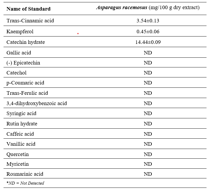
An HPLC method has been developed and validated for linearity, accuracy, stability, and precision to analyze sixteen polyphenolics simultaneously. After that, a methanolic extract of Asparagus racemosus was used to identify and quantify these polyphenolic compounds. Figure 1 illustrates the chromatographic separations of polyphenols in A. racemosus methanolic extract. A standard stock was prepared for the calibration curve, and the amount of each phenolic compound was extrapolated from its corresponding calibration curve. Catechin hydrate, Trans-cinnamic acid, and kaempferol were found in the methanolic extract of A. racemosus (Table 1).
Table 1: HPLC chromatogram of methanolic extract of Asparagus racemosus root


Figure 1: HPLC chromatogram of methanolic extract of Asparagus racemosus peaks: 1, Catechin hydrate; 2, Trans-cinnamic acid; and 3, Kaempferol.
HPLC-DAD analysis identified three major polyphenolic compounds in the methanolic extract: catechin hydrate (14.44 ± 0.09 mg/100 g), trans-cinnamic acid (3.54 ± 0.13 mg/100 g), and kaempferol (0.45 ± 0.06 mg/100 g). These compounds are known for their antioxidant and anti-inflammatory properties, which may contribute to the observed reno-protective effects
3.2 Effect of A. racemosus root extract on Renal Biomarkers in ISO induced rat
To evaluate the reno-protective potential of Asparagus racemosus root extract, key renal biomarkers were assessed across four experimental groups: Control, Control+ A. racemosus, ISO, and ISO + A. racemosus. The results are summarized in Figure 2 (Panels A–D).
Kidney Wet Weight has been shown in Figure 2A. ISO administration did not increase kidney wet weight (p > 0.05) significantly compared to control and control+ A. racemosus groups, indicating no evidence of renal hypertrophy and edema. Co-treatment with A. racemosus extract did not reduce kidney weight (p > 0.05 vs. ISO), suggesting not appreciable difference of kidney wet weight in ISO-induced rat model.
MPO Activity is demonstrated in Figure 2B. Myeloperoxidase (MPO) activity, a marker of neutrophil infiltration and inflammation, was significantly elevated in the ISO group (p < 0.01). Treatment with A. racemosus extract led to a significant reduction in MPO levels (p < 0.01 vs. ISO), indicating anti-inflammatory effects.
In Figure 2C, The Uric Acid Levels are represented. ISO-treated rats showed a significant rise in serum uric acid (p < 0.01), reflecting impaired renal clearance and oxidative stress. The ISO + A. racemosus group exhibited a significant decrease in uric acid levels (p < 0.05), approaching baseline values.
Plasma Creatinine depicted in Figure 2D. Creatinine levels were significantly elevated in the ISO group (p < 0.01), consistent with renal dysfunction. Co-administration of A. racemosus extract significantly lowered creatinine levels (p < 0.01 vs. ISO), suggesting improved renal filtration capacity.

Figure 2: Effect of A.racemosus root extract on kidney wet weight (A); MPO assessment (B); uric acid in Plasma (C); Creatinine in Plasma (D). All data were presented as mean ± SEM. For statistical analysis, one-way ANOVA followed by Tukey’s multiple comparison tests were done, where significant indicated as ns means p > 0.05; * means p ≤ 0.05; ** means p ≤ 0.01.
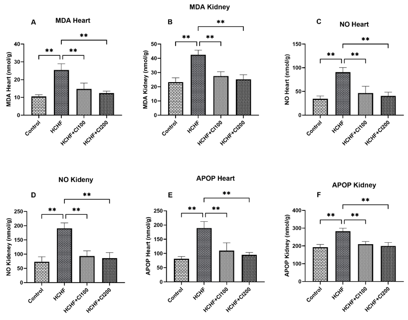
3.3 Effect of Asparagus racemosus Root Extract on Oxidative Stress Markers in Kidney Homogenates: MDA, NO, AOPP, and Catalase
MDA, NO, AOPP, and Catalase provide insight about the oxidative stress in an organ. The effect of Asparagus racemosus on oxidative stress in kidney has been evaluated. The result is shown in Figure 3.

Figure 3: Effect of A.racemosus root extract on oxidative stress markers MDA assessment (A); NO assessment (B); AOPP assessment (C); Catalase assessment (D). All data were presented as mean ± SEM. For statistical analysis, One way ANOVA followed by Tukey’s multiple comparison tests were done where significant indicated as ns means p > 0.05; * means p ≤ 0.05.
Malondialdehyde (MDA), a marker of lipid peroxidation showed in Figure 3(A). The ISO group exhibited a significant increase in kidney MDA levels (approximately 150 nmol/mL) compared to the control group (around 50 nmol/mL, p < 0.01). The group treated with ISO + A. racemosus had significantly lower MDA levels (around 75 nmol/mL) than the ISO group (p < 0.01), although they were still higher than the control group. The control + A. racemosus group showed no significant difference from the control group.
Figure 3(B) presents the concentration of nitric oxide (NO) in kidney tissue. Similar to the MDA results, the ISO group showed a significant elevation in NO levels (approximately 20 nmol/mL) compared to the control group (around 7.5 nmol/mL, p < 0.01). Treatment with A. racemosus (ISO + A. racemosus group) significantly reduced NO levels to approximately 10 nmol/mL compared to the ISO group (p < 0.01). The control + A. racemosus group was not significantly different from the control.
Advanced Oxidation Protein Products (AOPP), an indicator of protein damage displayed in Figure 3(C). The ISO group had markedly higher AOPP levels (approximately 1200 nmol/mL) than the control group (around 600 nmol/mL, p < 0.01). The ISO + A. racemosus group demonstrated a significant reduction in AOPP levels to around 700 nmol/mL when compared to the ISO group (p < 0.01). The control + A. racemosus group did not show a significant change from the control.
Antioxidant enzyme activity, Catalase illustrated in Figure 3(D). The ISO group showed a significant decrease in Catalase activity (approximately 15 U/min) compared to the Control group (around 30 U/min, p < 0.01). In contrast, the ISO + A. racemosus group showed a significant increase in Catalase activity (approximately 25 U/min) compared to the ISO group (p < 0.01). The Catalase activity in the Control + A. racemosus group was not significantly different from the Control.
3.4 Histopathological evaluation of A. racemosus on kidney in isoprenaline (ISO) administered rats

Figure 4: illustrates the histopathological changes in kidney tissue, focusing on inflammation and fibrosis, as a result of isoproterenol (ISO) administration and A. racemosus root extract treatment. The figure contains two panels of stained tissue images: hematoxylin and eosin (H&E) staining to show inflammatory cell infiltration (Panels A–D), and Sirius red staining to highlight collagen accumulation (Panels E–H).
3.4.1. Histological evaluation of Hematoxylin and Eosin staining of kidney section
The kidney tissue from the control group shows normal histological architecture with no signs of inflammatory cell infiltration. The ISO-treated group exhibits significant histological damage, marked by a substantial infiltration of inflammatory cells within the interstitial spaces of the kidney. The control group treated with A. racemosus shows a normal kidney structure, similar to the Control group, with no inflammation. The group treated with ISO and A. racemosus shows a notable reduction in inflammatory cell infiltration compared to the ISO-only group. The kidney structure appears much more preserved.
3.4.2. Histological evaluation of Sirius Red staining of kidney section
The lower panel (E–H) shows the Sirius red-stained kidney sections, which highlight collagen fibers in red. The Control group shows a normal distribution of collagen, with minimal red staining, indicating healthy kidney tissue. The ISO-treated group displays a pronounced increase in collagen deposition, evidenced by extensive red staining, which indicates significant fibrosis. The Control group treated with A. racemosus shows no change in collagen distribution compared to the Control, indicating no fibrotic effects. The group treated with ISO and A. racemosus shows a clear decrease in collagen accumulation compared to the ISO-only group, suggesting a reduction in fibrosis.
4. Discussion
5. Limitations And Future Perspectives
6. Conclusion
Author Contribution
Funding
Institutional Review Board Statement
Data Availability Statement
Acknowledgments
Conflicts of Interest
References
Figures