Original article Open access | J. Bio. Exp. Pharm. 2025, 3(1), 01-19 | https://doi.org/10.62624/JBEP00.022
Evaluation of Coccinia grandis Extract on Cardiac and Kidney Dysfunction in High-Carbohydrate High-Fat Diet-Fed Rats
Shahnaz Siddiqua, Sungida Sharmin, Tanjila Jalal, Faiza Hamid Jyoti, Mehzabin Rahman, Md. Tareq Chowdhury, Nabila Alam, Karima Boshra, Nusrat Subhan, Md Ashraful Alam

Abstract

Obesity is a chronic metabolic disorder that is closely related to cardiovascular and renal dysfunctions due to oxidative stress, inflammation, and lipid abnormalities. This study aimed to investigate the cardioprotective and renoprotective effects of ethanolic leaf extract of Coccinia grandis (CI) on high carbohydrate, high fat (HCHF) diet-induced obese Wistar rats. Twenty-four male rats were divided into four groups: Control, HCHF, HCHF + CI100 (100 mg/kg), and HCHF + CI200 (200 mg/kg), and treated orally for 56 days. Biochemical, oxidative stress, and antioxidant enzyme parameters were evaluated in plasma, heart, and kidney tissues, followed by histological analysis. The HCHF diet significantly increased body weight and organ weights, LDL, CK-MB, uric acid, creatinine, MDA, MPO, and NO levels, while decreasing catalase and SOD activity. Treatment with C. grandis extract at both doses significantly reversed these alterations by restoring antioxidant enzyme activity and reducing lipid peroxidation and inflammatory markers. Histological studies showed a marked decrease in fibrosis, collagen deposition, and inflammatory cell infiltration in cardiac and renal tissues of C. grandis-treated groups, with higher doses providing greater protection. These protective effects are attributed to the antioxidant and phenolic constituents of C. grandis, which counter oxidative stress-mediated damage. Overall, C. grandis ethanolic extract demonstrated potent therapeutic potential against diet-induced cardiorenal injury by improving lipid metabolism, oxidative balance, and tissue integrity. These findings suggest the promising application of C. grandis in the management of metabolic syndrome and obesity-related complications.


Keywords

Coccinia grandis
Oxidative stress
Antioxidant enzymes
Cardiovascular disease
Renal injury

1. Introduction

 Obesity is a chronic and complicated illness that can lower mental, social, and physical health, ultimately having an impact on the quality of life [1]. A high body mass index (BMI) or waist circumference is typically used to measure obesity, or more generally, being overweight, and it leads to numerous issues [2]. The two primary causes of health problems are the toxic chemicals released by larger fat cells and the increased bulk of fat tissue. Physical mass causes issues such as joint injury, sleep apnea, and social shame. At the same time, the metabolic impacts of fat cells lead to diseases like insulin resistance, type 2 diabetes, inflammation, blood clotting problems, heart disease, and some types of cancer [3]. Larger gastrointestinal tracts cause people to store more subcutaneous fat, and leptin deficiency shows that genetics may be a strong regulator of body weight [4-6]. Obesity and heart failure (HF) are both complicated conditions that frequently have connections. In the United States (US), 6.7 million adults suffer from heart failure (HF), and by 2030, that number is predicted to increase to 8.5 million [7]. Oxidative stress (OS) is one of the most important pathogenic processes of myocardial infarction (MI). It is a complicated system network that includes mitochondrial malfunction, inflammatory response, and excess calcium [8]. When too much reactive oxygen species (ROS) exceeds the antioxidant capacity of the cell, cardiovascular problems such as endothelial dysfunction, atherosclerosis, ischemia/reperfusion (I/R) damage, etc., are caused [9]. This excess ROS can cause inflammation to worsen and initiate a vicious loop that draws leukocytes to the reperfusion region and causes myocardial cell necrosis and death [10]. The pathophysiology of cardiovascular disease (CVD) is known to be significantly influenced by elevated oxidative stress and diminished antioxidant defense. It has been noted that MI patients have lower levels of catalase and SOD activity [11]. Maintaining cellular homeostasis depends on lipid metabolism, an essential physiological function. However, excess waste, particularly lipid peroxides, which are closely associated with the development and progression of acute myocardial infarction (AMI), accumulates when this metabolic pathway is disturbed [12]. Recent research has shown that metabolic syndrome (MS), overweight, and obesity are powerful independent risk factors for end-stage renal disease (ESRD) and chronic kidney disease (CKD) [13]. Proteinuria, lipid accumulation, fibrosis, and glomerulosclerosis are all symptoms of obesity-related chronic kidney disease, and macrophages are a major factor in causing inflammation and kidney damage [14].
The climbing tropical perennial vine Coccinia grandis (CI), also referred to as ivy gourd, is a member of the Cucurbitaceae family and is indigenous to the tropical regions of North and Central Africa as well as Southeast Asia [15]. In traditional Indian medicine, the leaves of this plant are used to lower the blood sugar levels of diabetic patients. Triterpenoid, flavonoid, lupeol, β-amyrin, β-sitosterol, B-β-carotene, lycopene, and cryptoxanthin are all found in the plant's root and stem. Antibacterial, anthelmintic, antimalarial, anti-inflammatory, antioxidant, antipyretic, analgesic, antiulcer, hypoglycemic, antifungal, antitussive, mutagenic, and alpha-amylase-inhibiting effects are only a few of the pharmacological properties that this plant has demonstrated [16, 17]. Its antifibrotic and antioxidant properties, which are mediated by bioactive compounds such flavonoids, phenols, and glycosides imply that it may be useful as a treatment for diseases caused by inflammation and oxidative stress [18, 19]. Its antioxidant qualities are thought to help prevent kidney damage and regulate lipid peroxidation, which would exacerbate oxidative damage in diabetes caused by streptozotocin [20, 21].

2. Materials & Method

2.1 Identification and collection of plant materials
We collected leaves of Coccinia grandis from the Cumilla district. The sample's validity was confirmed by Bangladesh's National Herbarium. For future reference, a specific voucher (DABC Accession Number: 47416) is collected for Coccinia grandis (CI).
 
2.2 Plant process
Coccinia grandis had to be properly rinsed with water before being left to dry naturally. The withering leaves were finally ground into a fine powder. For fifteen days, the powder was submerged in 70% ethanol. The conical flask was covered with aluminum foil and placed in an incubator shaker at 180 rpm at 37 °C for 1 week. Next, Whatman No. 1 filter paper was used to filter the plant extract. The extract was then condensed at low temperature (32–40 °C) by evaporation using a rotary evaporator. The glassware was cleaned and rinsed with deionized water and kept at 160 °C for 2 h. Plasticware was autoclaved before initiation of the antimicrobial experiment [22]. Finally, the raw materials were sent for the required pharmacological analysis [17].
 
2.3 Experimental design
From the animal breeding facility of North South University, 24 males Wistar Rats were collected, weighing around 185–200 g, between the ages of ten and twelve weeks. Separate cages were provided for the rats and they were kept at a controlled temperature of 22±3 ⁰C where the light and day cycles were 12 hours apart. All the rats had free access to food and water. Experimental protocols were accepted from the Ethical Committee of North South University (AEC 009-2018). The rats were divided into groups of four, with each group consisting of 6 rats.
Group I- Control group, which received normal food and water for 56 days.
Group II- High carbohydrate high fat (HCHF) group which also took for 56 days.
Group III- HCHF+CI100 which took both high high-carbohydrate high-fat diet and 100 mg/kg leaves ethanol extract of Coccinia grandis (CI) by oral gavage for 56 days.
Group IV- HCHF+CI200 which took both high carbohydrate high fat diet and 200 mg/kg leaves ethanol extract of Coccinia grandis (CI) by oral gavage for 56 days.
The body weight, food intake and water intake were recorded for 8 weeks and at the 57th day Ketamine (90 mg/kg) was used to sacrifice the rats for further blood and organ collection. Blood was collected in 1.5 cc micro centrifuge tube by a 5cc syringe where citrate buffer was kept as anticoagulant agent from large abdominal vein. After that blood was taken for centrifuge system where blood was centrifuged at 4000 rpm at 4 ⁰C for 15 minutes. After plasma were separated and were kept at -20 ⁰C for biochemical assay. Different types of organs were also collected such as heart and kidney which were divided into two parts such as for biochemical assay and for histological analysis. Organs which were kept for biochemical assay were kept in -18⁰C and histological purpose organs were kept in neutral buffered formalin which pH was 7.4.
High Carbohydrate High Fat diet was prepared in pellet form in the pharmacology laboratory of North South University (Table 1).
 
2.4 Plasma Biochemistry analysis
LDL, CK-MB, Uric Acid, and Creatinine tests were all performed according to the manufacturer protocol provided by Diatec Diagnostic Ltd (Hungary).
 Table 1: Control diet and high-carbohydrate high-fat diet composition 
2.5 Tissue sample preparation and oxidative stress parameters assay
For analysis, Heart and kidney tissues were selected. Tissue was homogenized in phosphate buffer solution where pH was around 7.4 and volume was 10 mL. The centrifugation parameter was 8000 rpm at 4 ⁰C for 15 minutes [23]. After centrifugation, supernatants were collected from tissue homogenates.
 
2.5.1 Malondialdehyde (MDA) assay
An MDA standard was used to establish a standard curve against which the sample readings were plotted. This method depends on the formation of MDA as a byproduct of lipid peroxidation, which reacts with thiobarbituric acid to produce thiobarbituric acid reactive substance (TBARS), a pink chromogen that can be measured spectrophotometrically at 532 nm [24]. In our study we identify the MDA concentration of Heart and kidney. Heart and kidney tissue samples were diluted in Phosphate buffered saline in an Eppendorf. Then 100 µL glacial acetic acid was added and the sample was let to rest for 15 minutes. After that, we added 200 µL TBA (0.37%) and sealed the Eppendorf tube. The sealed Eppendorf kept in hot water bath for 10 minutes. Then the mixture was cool in room temperature, 200 µL of sample take in 96 well plate. Take two times of absorbance at 490 nm.
 
2.5.2 Nitric oxide (NO) assay
To measure the generation of nitric oxide, Griess reagent was used [25]. 20 µL of heart and tissue samples were dissolved in 80 µL PBS and then 50 µL sulfanilamide was added. After an interval of 5 minutes, 50 µL of 0.1% w/v naphthyl ethylene diamine dihydrochloride (NED) were then added to the sample mixture and again after an interval of 10 minutes we took the absorbance at 540 nm [26]. NaNO2 was used as a standard to make the standard curve, and the units were represented as mmol/ml or mmol/g of tissue.
 
2.5.3 Advanced oxidation protein products (APOP) assay
In this process 10 µL of sample was taken in 90 µL PBS in a 96 well plate. 50 µL glacial acetic acid (15%) was used to turn the media into acidic. To create a colored complex, 50 µL of potassium iodide (1.16 mM) was then added. The absorbance at 405 nm was measured after two minutes of waiting. Chloramine T was used as a standard, and units were calculated as mmol/mL or mmol/g of tissue.
 
2.6 Antioxidant enzyme activity assay
Superoxide dismutase (SOD) activity was determined by the nitro blue tetrazolium technique, which is based on the decreased activity of NBT that SOD reduces [25]. To determine SOD activity of heart and kidney tissue were selected. 10µL sample of heart and kidney tissue dissolved in 90µL PBS in a 96-well plate. Then added 100 µL adrenaline injection. Finally, absorbance is taken in 0 min,1 min, 2min, 3 min, 4 min at 490 nm.
 
2.7 Myeloperoxidase (MPO) activity estimation
10 µL tissue of heart and kidney dissolved in 90 µL PBS in a 96-well plate. Then added 3µL of H2O2 (0.15 mM). After that, 3µL of o-dianisidine solution was added. The absorbance was taken at 460 nm and reported as MPO activity/mg protein.
 
2.8 Histological Study
In case of histology heart and kidney tissue were fixed in neutral buffered formalin. To embed in paraffin wax serial xylene and alcohol treatment were done. In a rotary microtome sections were about 5 μm for each tissue block [23]. In heart and kidney tissue, Hematoxylin and Eosin stain was used for the determination of inflammatory cell infiltration. For determination of collagen deposition Sirius red staining was done for both heart and kidney tissue. To capture the pictures of histology a light microscope (Zeiss Axioscope) was used and all the pictures were taken at 40x magnifications.
 
2.9 Statistical analysis
To measure all data mean ± SEM were used to represent all values. For statistical significance one way ANOVA Newman-Kuels post hoc test were selected to perform comparison between groups. All results were prepared by using GraphPad Prism Software (version 9). In all cases statistical significance were considered as p<0.05                                
 

3. Result

3.1 Effect of Coccinia grandis (CI) on body weight, food intake and water intake in high-carbohydrate high-fat (HCHF) diet induced rats
In the body weight HCHF group increased a large amount of body weight compared to control group (Figure 1A). On the other hand, both HCHF+CI100 and HCHF+CI200 reduced body weight compared to HCHF group (Figure 1A). In case food intake HCHF group consumed a much higher amount food compared to control group (Figure 1B). On the other hand, no significant difference was found in the food amount consumed by both HCHF+CI100 and HCHF+CI200 when compared with the HCHF group (Figure 1B). In case of water intake, no significant difference was found between any groups (Figure 1C).
 
Figure 1: Effect of Coccinia grandis (CI) on body weight, food intake and water intake in High Carbohydrate High Fat (HCHF) diet administered rats. Here 1A: Body weight; 1B: Food intake and 1C: Water intake. For statistical analysis one-way ANOVA and Newman-Keuls post hoc test were followed. In each group 6 rats were taken which means n=6. Every data was calculated by Mean ± SEM. Significance of value were considered at p<0.05.
 
3.2 Effect of Coccinia grandis (CI) on total heart wet weight and kidneys wet weight in High Carbohydrate High Fat (HCHF) diet-treated rats
In case of groups with received HCHF diet, the total heart wet weight increased significantly (p≤0.01) compared to the control group (Figure 2A). On the other hand, rats which took 100 mg/kg ethanolic extract of Coccinia grandis by oral gavage and HCHF diet, their total heart wet weight was reduced significantly (p≤0.01) when compared with the rats of the group which received only the HCHF diet (Figure 2A). Additionally, it was seen that the HCHF + CI200 (where the rats were fed with 200 mg/kg ethanolic extract of Coccinia grandis) group also reduced the total heart wet weight significantly (p≤0.01).
The kidney wet weights across various groups also had seen fluctuations in the figure 2B, the group treated with only HCHF had a significantly increased kidney wet weight when compared to the control group. Interestingly, both our treatment groups at different doses, HCHF + CI100 and HCHF + CI200, had remarkably reduced this increased kidney wet weight seen in the disease group fed with only HCHF diet.
 
Figure 2: Effect of Coccinia grandis (CI) on total heart wet weight and kidneys wet weight in High Carbohydrate High Fat (HCHF) diet induced rats. Here 2A: Total Heart Wet Weight; 2B: Kidneys Wet Weight. For statistical analysis one-way ANOVA Newman-Keuls post hoc test were followed. In each group 6 rats were taken which means n=6. Every data was calculated by Mean ± SEM. Significance of value were considered at p<0.05.
 
3.3 Effect of Coccinia grandis (CI) on LDL plasma, MPO heart and MPO kidney in High Carbohydrate High Fat (HCHF) diet administered rats
Figure 3: Effect of Coccinia grandis (CI) on low density lipoprotein, myeloperoxidase heart and myeloperoxidase kidney in High Carbohydrate High Fat (HCHF) diet treated rats. Here 3A: LDL; 3B: MPO Heart; 3C: MPO Kidney. For statistical analysis one-way ANOVA Newman-Keuls post hoc test were followed. In each group 6 rats were taken which means n=6. Every data was calculated by Mean ± SEM. Significance of value were considered at p<0.05.
 
In figure 3A it can be seen that the LDL concentration of plasma in the HCHF group had increased significantly when compared with the control group, In case of the rats which were treated with 2 different doses of our ethanolic extract of Coccinia grandis, it was seen that the treatments at both doses could significantly restore the LDL levels at a level similar to the control group.
In figure 3B the supplementation of HCHF group throughout the trial showed a drastic increase in the MPO levels of heart when compared with the control groups, our treatment groups on the other hand HCHF+CI100 decreased MPO heart activity significantly (p≤0.01) compared to HCHF group. HCHF+CI200 group also decreased MPO heart activity significantly (p≤0.01) compared to HCHF group.
In figure 3C, it was seen that due to the rats only being fed HCHF diet, the kidney tissues were also affected and had an increase in the MPO levels when compared to the control group. Both our treatment doses showed significant reduction in the amount of MPO activity when compared to the only HCHF fed groups.
 
3.4 Effect of Coccinia grandis (CI) on oxidative stress parameters of heart and kidney in High Carbohydrate High Fat (HCHF) diet administered rats
 
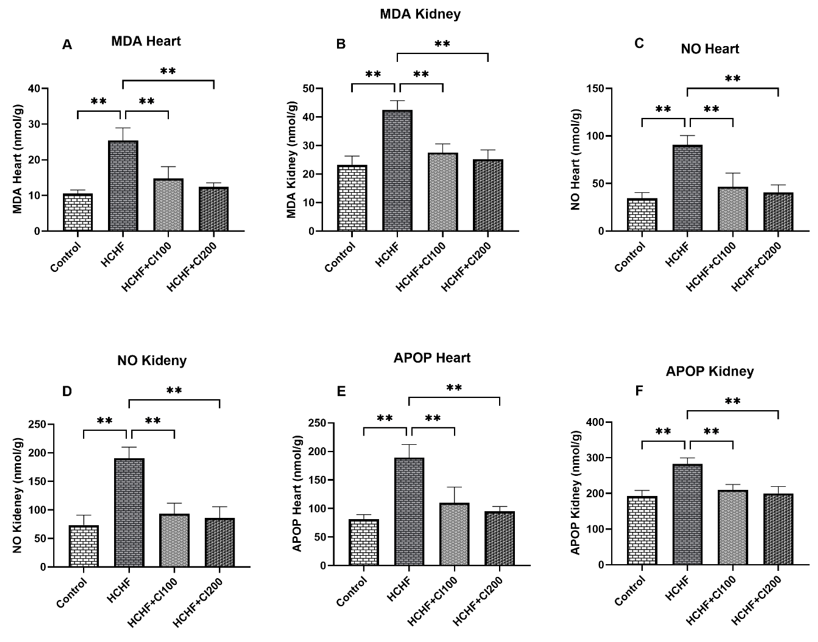
Figure 4: Effect of Coccinia grandis (CI) on oxidative stress parameters of heart and kidney in High Carbohydrate High Fat (HCHF) diet treated rats. Here 4A: MDA Heart; 4B: MDA Kidney; 4C: NO Heart; 4D: NO Kidney; 4E: APOP Heart; 4F: APOP Kidney. For statistical analysis one-way ANOVA Newman-Keuls post hoc test were followed. In each group 6 rats were taken which means n=6. Every data were calculated by Mean ± SEM. Significance of value were considered at p<0.05.
 
MDA and NO analysis showed that both markers had significantly increased in both heart and kidney tissues rapidly in the group which was only fed with HCHF diet, when compared with the control group. The two groups treated with two different doses of our ethanolic extract (CI 100, CI 200) on the rats fed with HCHF diet, showed much restoration in the MDA and NO concentrations for both organs compared to the obese group (Figure 4A, 4B, 4C, 4D). Interestingly, it was seen that for both cardiac and renal analysis of MDA and NO, the HCHF + CI200 group worked a bit better than the HCHF + CI100 group in reducing the oxidative stress marker levels.
AOPP concentration for both renal and cardiac tissues were significantly elevated in the HCHF group when compared with the control group (p≤0.01) (Figure 4F) and this elevation was decreased greatly in the HCHF + CI100 and HCHF + CI200 groups. Similarly to previously found results for MDA and NO oxidative stress markers, Coccinia grandis ethanolic extract at 200 mg/kg supplemented group (HCHF + CI200) worked slightly better than the 100 mg/kg supplemented treatment group (HCHF + CI100).
 
3.5 Effect of Coccinia grandis (CI) on antioxidant enzyme activity parameters of heart and kidney in High Carbohydrate High Fat (HCHF) diet administered rats
 
Figure 5: Effect of Coccinia grandis (CI) on antioxidant enzyme activity parameters of heart and kidney in High Carbohydrate High Fat (HCHF) diet treated rats. Here 5A: Catalase Heart; 5B: Catalase Kidney; 5C: SOD Heart; 5D: SOD Kidney. For statistical analysis one way ANOVA Newman-Keuls post hoc test were followed. In each group 6 rats were taken which means n=6. Every data were calculated by Mean ± SEM. Significance of value were considered at p<0.05.
 
HCHF administration significantly reduced the activity of antioxidant enzymes (SOD & Catalase) in both cardiac and renal tissues when compared to the control group (Figure 5).
The catalase activity was significantly restored in cardiac and renal tissues in both groups (HCHF + CI100 & HCHF + CI200) i.e. both 100 mg/kg and 200 mg/kg doses of CI significantly restored the decreased catalase enzyme activity.
Similarly, the HCHF induced reduction in SOD enzyme activity was also significantly reversed due to the administration of both CI doses in both cardiac and renal tissues. Interestingly, for the restoration of antioxidant enzyme activities in both cardiac and renal tissues, the 200 mg/kg dose of CI worked better than the 100 mg/kg dose of CI.
 
3.6 Effect of Coccinia grandis (CI) on CK-MB plasma, Uric Acid plasma and Creatinine plasma in High Carbohydrate High Fat (HCHF) diet induced rats
 
Figure 6: Effect of Coccinia grandis (CI) on CK-MB plasma, Uric Acid plasma and Creatinine plasma in High Carbohydrate High Fat (HCHF) diet fed rats. Here 6A: CK-MB plasma; 6B: Uric Acid plasma; 6C: Creatinine Plasma. For statistical analysis one way ANOVA Newman-Keuls post hoc test were followed. In each group 6 rats were taken which means n=6. All data were calculated by Mean ± SEM. Significance of value were considered at p<0.05.
 
In Figure 6, it was seen that the HCHF diet significantly increased the concentrations of cardiac and renal biomarkers, such as CK-MB, Uric Acid, and creatinine levels in plasma, when compared with the control group.
The plasma concentration of CK-MB, an enzyme primarily found in the heart muscle released to the blood stream during cardiac damage (i.e. a marker for cardiac injury), was highly elevated in the HCHF group when compared to the control group and the treatment with C. grandis ethanolic extract in groups treated with both 100 mg/kg and 200 mg/kg doses (HCHF + CI100 & HCHF + CI200), significantly restored the elevated plasma levels compared to the HCHF only group (Figure 6A).
Uric acid levels in plasma (a renal biomarker) were also significantly increased in the HCHF group. Both CI treatment doses significantly reduced the uric acid concentration when compared to the only HCHF fed group. (Figure 6B).
Plasma creatinine levels, another important renal function marker, were also significantly increased in the HCHF group compared to the control group and the administration of CI at both 100 mg/kg and 200 mg/kg concentrations significantly decreased the concentration of creatinine in plasma compared to the HCHF group. (Figure 6C).
In cases of all the cardiac and renal damage markers, CK-MB, Uric Acid and Creatinine, the 200 mg/kg dose of CI worked slightly better in reducing or restoring the biomarker level when compared to the 100 mg/kg dose of CI.
 
3.7 Effect of Coccinia grandis (CI) on histopathological study in High Carbohydrate High Fat (HCHF) diet induced rats
 
 
Figure 7: Effect of Coccinia grandis (CI) on hematoxylin and eosin staining of heart in High Carbohydrate High Fat (HCHF) diet fed rats. Here 7A: Control; 7B: HCHF; 7C: HCHF+CI100; 7D: HCHF+CI200. All pictures were taken at 40X magnification.
Control group showed no inflammatory cell infiltration or any kind of abnormalities of heart (Figure 7A). HCHF group showed a major cardiac fibrosis and additionally cell infiltration (Figure 7B). The group which took 100 mg/kg ethanol extract of Coccinia grandis (CI) in high carbohydrate high fat induced rats showed a decent amount of recovery from cell infiltration in cardiac tissue (Figure 7C). Group which belongs to HCHF+CI200 showed much improved cardiac cell condition compared to HCHF group (Figure 7D).
 
 
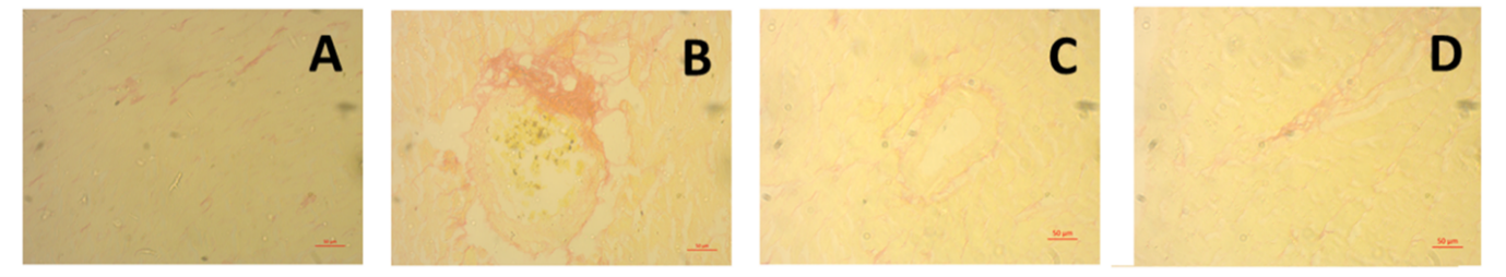
Figure 8: Effect of Coccinia grandis (CI) on Sirius red staining of heart in High Carbohydrate High Fat (HCHF) diet fed rats. Here 8A: Control; 8B: HCHF; 8C: HCHF+CI100; 8D: HCHF+CI200. All pictures were taken at 40X magnification.
Control group did not show any kind of collagen deposition and fibrosis which means it is normal in heart (Figure 8A). HCHF group showed a huge amount of fibrosis and collagen deposition in cardiac cell (Figure 8B). Collagen deposition was reduced in HCHF+CI100 group which means it actually protects cardiac muscle (Figure 8C). HCHF+CI200 also showed a well recovery from collagen deposition which also means heart tissue is in quite good condition (Figure 8D).
 
 
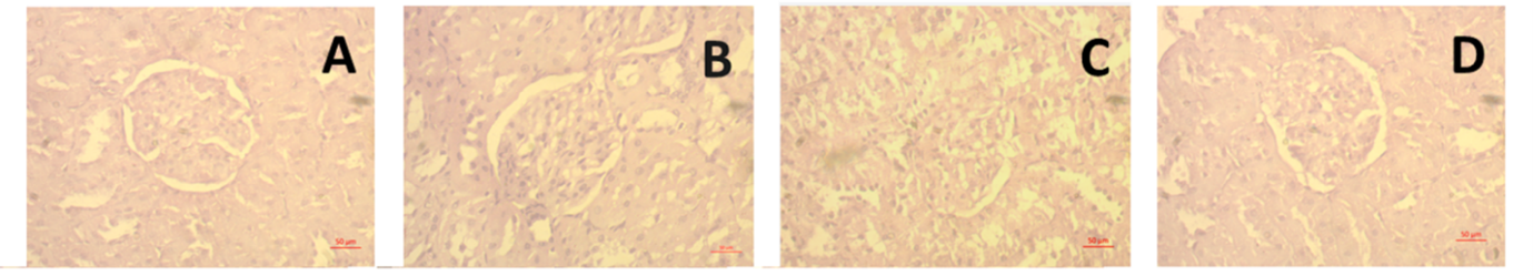
Figure 9: Effect of Coccinia grandis (CI) on hematoxylin and eosin staining of kidney in High Carbohydrate High Fat (HCHF) diet induced rats. Here 9A: Control; 9B: HCHF; 9C: HCHF+CI100; 9D: HCHF+CI200. All pictures were taken at 40X magnification.
Control group showed no cell infiltration or any kind of abnormalities in kidney (Figure 9A). HCHF group showed a major cell infiltration in kidney (Figure 9B). The group which was treated with 100 mg/kg ethanol extract of Coccinia grandis (CI) in high carbohydrate high fat administered rats showed recovery from cell infiltration in kidney tissue (Figure 9C). Group which belongs to HCHF+CI200 showed much improved kidney tissue condition compared to HCHF group (Figure 9D).
 
 
Figure 10: Effect of Coccinia grandis (CI) on Sirius red staining of kidney in High Carbohydrate High Fat (HCHF) diet fed rats. Here 10A: Control; 10B: HCHF; 10C: HCHF+CI100; 10D: HCHF+CI200. All pictures were taken at 40X magnification.
Control group did not show any kind of collagen deposition which means it is normal in kidney (Figure 10A). HCHF group showed collagen deposition in kidney cell (Figure 10B). Collagen deposition was reduced in HCHF+CI100 group which means it actually protects renal tissue (Figure 10C). HCHF+CI200 also showed a well recovery from collagen deposition which also means kidney tissue can be protected by Coccinia grandis (CI) (Figure 10D).

4. Discussion

In addition to a variety of metabolic risk factors, obesity is a distinct risk factor for CVD [27]. According to estimates, obesity's impact on metabolic risk factor levels contributes to half of the risk of CVD disease, with remaining percentage most likely caused by separate causes. Monosodium glutamate in high-lipid diets (MH) leads to common health problems including chronic kidney disease (CKD) and renal fibrosis through oxidative stress and inflammation [28]. In rats fed with a HF diet, C. grandis treatment greatly lowered the buildup of peritoneal and epididymal fat, liver excess weight, and cholesterol levels. According to the results for potential antioxidants, obesity and dyslipidemia were effectively reduced with doses of C. grandis extracts [19].
 
To determine if C. grandis is effective in lowering lipid peroxidation, LDL, and collagen accumulation which protect renal tissue recovery and cell infiltration, fibrosis, and repairing kidney tissue, an in-depth meta-analysis and review was carried out. Our research shows that a diet heavy in fat and carbohydrates raises LDL. In patients it has been shown that elevated oxidized LDL levels have been linked to structural change, cardiac dysfunction [29]. When aqueous leaf extract of C. grandis was administered to diabetic mice induced by alloxan, Manjula and Ragavan reported decreases in blood glucose, TC, TG, VLDL, and LDL [30]. Coccinia grandis extract significantly reduced LDL in a rat model which was fed a high-fat diet, according to another study [17].
In this study it was observed that both doses, 100 mg/kg and 200 mg/kg of Coccinia grandis (CI) decreased the LDL concentration.
 
A recognized way to cause obesity and insulin resistance in rodents is to feed them a high-fat diet (HFD). Excessive adipose tissue deposit encourages immune cells, such as macrophages, to infiltrate the body and produce reactive oxygen species (ROS) and pro-inflammatory cytokines. An imbalance of redox equilibrium can be affected by these increased ROS, which can cause inflammation and cellular damage, ultimately contributing to the course of issues linked to obesity [31]. The HFD raises oxidative stress and NF-κB function, activates macrophages, and increases endotoxins and free fatty acids [32]. The main source of oxidants that alter and change LDL into a high-uptake form that macrophage scavenger receptors can recognize is MPO [33-36]. This results in macrophage foam cells, which are crucial in increasing the formation of atherosclerosis.
 
Higher levels of oxidative stress caused by a high-fat diet is one factor that leads to obesity's metabolic syndrome. But since Coccinia grandis contains antioxidants like quercetin, it reduces this stress [16] . According to this study, a high-carb, high-fat diet increased the level of several oxidative stress parameters in the heart and kidney tissue. Ethanol extract of (CI) leaf at two different doses, such as 100 mg/kg and 200 mg/kg, reduced these levels.
 
One of the primary causes of acute kidney injury is renal ischemia-reperfusion (IR) injury, which is affected by the duration of the ischemia and any presence of comorbidities [37]. Eating a high-fat diet has been observed to cause metabolic dyshomeostasis and renal lipotoxicity, which may affect the kidney's essential processes [25]. Rats from both the ND and HD groups that were exposed to IR, however, showed reduced GSH/GSSG levels along with significantly lower antioxidant enzyme activity in SOD. Endogenous antioxidant enzymes like SOD and CAT protect human and animal cells from FR damage. The body generally defends itself against ROS through a form of self-defense. This system is referred to as an anti-oxidative system, and it can be either non-enzymatic (GSH) or enzymatic (SOD, CAT, and GPx).
 
The amount of ROS created, however, may be greater than what the body's antioxidants are capable of controlling when hyperglycemia is present. Poor cell metabolism results from changes in the redox signal of cells caused by an imbalance in the ROS: antioxidant ratio [38].By decreasing renal TBARS, pioglitazone or leaf extract from Coccinia grandis slowly decreases stress brought on by reactive oxygen species with diabetes-induced renal damage. These methods also increased the concentrations of SOD and catalase at the same time [21]. Two tests have been performed in this experiment to measure the antioxidant enzyme activity, including catalase and SOD. Our findings show that administering Coccinia grandis (CI) leaf ethanol extract can either increase or restore antioxidant enzyme activity, meaning that catalase and SOD enzyme activity continued to exist in the kidney and heart.
 
Both CK-MB and troponin-T levels had been significantly increased after consuming HFD [39]. According to the author, Wistar rats given a high-fat, high-cholesterol diet displayed higher levels of CK-MB, which is linked to functional and structural changes in the heart muscle and a breakdown of the cell membrane's functionality. This condition may also be due to lipid peroxidation in the heart membranes. identical results were reported by Wang et al., who compared the inflammatory action of saturated fatty acids to the rise in CK-MB and LDH [40, 41].
Additionally, the HM group showed a notable increase in cardiac markers LDH, CK-MB, Troponin I, in regard to the NC group. Following EECGL injections, the EECGLH group's levels of LDH, CK-MB, Troponin I, and T were significantly lower than those of the HM group [42]. Our research results showed that a high-carb, high-fat diet caused an increase in plasma CK-MB levels. The use of Coccinia grandis (CI) leaf ethanol extract as a treatment actually reduced the concentration of CK-MB, showing that it can be highly significant in cases of CK-MB.
In some studies, rats fed a high-fat diet (HFD) may develop chronic kidney injury. Clinical indications of this injury include glomerular shrinkage, mesangial cell and matrix development, ambiguous renal tubule shape, fibrosis, and persistent inflammation [43]. Serum creatinine, blood urea nitrogen, and urine protein synthesis all dropped in rats with diabetes-induced nephropathy when Coccinia grandis leaf extract and a low dose of pioglitazone were given together [21]. According to our research, Coccinia grandis (CI) may reduce creatinine at both low and high dosages.
Hematoxylin and eosin and Sirius red were two ways of staining used in the histology of the kidney and heart. Hematoxylin and eosin staining by a high-carb, high-fat diet show fibrosis and infiltration of heart and kidney cells in both cases. In the contrary, a high-carb, high-fat diet results in collagen accumulation in Sirius red staining for the kidneys and heart. At doses of roughly 100 mg/kg and 200 mg/kg, Coccinia grandis (CI) leaf ethanol extract stopped cell infiltration, fibrosis, and collagen deposition. Finally, we can say that these data confirm our hypothesis that diet-induced obesity can be controlled by Coccinia grandis.

5. Conclusion

In this study it is revealed that in high carbohydrate high fat diet ethanol extract of Coccinia grandis (CI) was used which actually retained antioxidant activity. As high carbohydrate high fat diet was administered it actually caused cardiac fibrosis and renal damage which was restored by the ethanolic extract of Coccinia grandis (CI), consisting of phenolic compounds. With the treatment of Coccinia grandis (CI) ethanolic extract, antioxidant enzyme activity was improved in catalase and SOD. Although the specific chemical compounds present in Coccinia grandis responsible for these protective effects were not identified, our findings can suggest that it possesses notable therapeutic potential against oxidative damage associated with metabolic syndrome and hypercholesterolemia, opening promising windows for future clinical applications for our plant in the management of metabolic and cardiovascular disorders.

Contrary to popular belief, Lorem Ipsum is not simply random text. It has roots in a piece of classical Latin literature from 45 BC, making it over 2000 years old. Richard McClintock, a Latin professor at Hampden-Sydney College in Virginia, looked up one of the more obscure Latin words, consectetur, from a Lorem Ipsum passage, and going through the cites of the word in classical literature, discovered the undoubtable source. Lorem Ipsum comes from sections 1.10.32 and 1.10.33 of "de Finibus Bonorum et Malorum" (The Extremes of Good and Evil) by Cicero, written in

Author Contribution

Conceptualization, MAA and NS; methodology, SS, SS and NA; software, MTC and KB; validation, MR, FHJ, and TJ; formal analysis, MR, MAA, and NS; investigation, SS, SS, MTC, NA and KB; resources, MR, NA and KB; data curation, MAA and NS; writing—original draft preparation, SS, TJ, MR, KB; writing—review and editing, FHJ, MTC, NA, KB; visualization, MAA, NS; supervision, MAA and NS project administration, MAA; funding acquisition, MAA. All authors have read and agreed to the published version of the manuscript.

Funding

 This research did not receive any internal or external funding from profit and non-profit organization. 

Institutional Review Board Statement

Not Applicable (N/A)

Data Availability Statement

Data used in this study will be available upon reasonable request from the corresponding author.

Acknowledgments

Authors are gratefully acknowledging the logistic support from the department of Pharmaceutical Sciences, North South University.

Conflicts of Interest

The authors declare no conflict of interest. The funders had no role in the design of the study; in the collection, analyses, or interpretation of data; in the writing of the manuscript, or in the decision to publish the results.

References

  1. Melson, E.; Ashraf, U.; Papamargaritis, D.; and Davies, M.J. What is the pipeline for future medications for obesity? Int. J. Obes. 2025, 49(3), 433-451. https://doi.org/10.1038/s41366-024-01473-y
  2. Bovet, P.; Farpour-Lambert, N.; Banatvala, N.; and Baur, L. Obesity: Burden, epidemiology and priority interventions. In Noncommunicable Diseases; Routledge: 2023; pp 74-82. https://doi.org/10.4324/9781003306689-12
  3. Bray, G.A. Medical Consequences of Obesity. J. Clin. Endocrinol. Metab. 2004, 89(6), 2583-2589. https://doi.org/10.1210/jc.2004-0535
  4. Budnik, A.; Henneberg, M. Worldwide Increase of Obesity Is Related to the Reduced Opportunity for Natural Selection. PLoS One 2017, 12(1), e0170098. https://doi.org/10.1371/journal.pone.0170098
  5. Henneberg, M.; Ulijaszek, S.J. Body frame dimensions are related to obesity and fatness: Lean trunk size, skinfolds, and body mass index. Am. J. Hum. Biol. 2010, 22(1), 83-91. https://doi.org/10.1002/ajhb.20957
  6. Lucas, T.; Henneberg, M. Body frame variation and adiposity in development, a mixed-longitudinal study of "Cape Coloured" children. Am. J. Hum. Biol. 2014, 26(2), 151-5. https://doi.org/10.1002/ajhb.22494
  7. Bozkurt, B.; Ahmad, T.; Alexander, K.M.; Baker, W.L.; Bosak, K.; Breathett, K.; Fonarow, G.C.; Heidenreich, P.; Ho, J.E.; Hsich, E.; Ibrahim, N.E.; Jones, L.M.; Khan, S.S.; Khazanie, P.; Koelling, T.; Krumholz, H.M.; Khush, K.K.; Lee, C.; Morris, A.A.; Page, R.L. II; Pandey, A.; Piano, M.R.; Stehlik, J.; Stevenson, L.W.; Teerlink, J.R.; Vaduganathan, M.; and Ziaeian, B. Heart Failure Epidemiology and Outcomes Statistics: A Report of the Heart Failure Society of America. J. Card. Fail. 2023, 29(10), 1412-1451. https://doi.org/10.1016/j.cardfail.2023.07.006
  8. Laule, C.; Rahmouni, K. Leptin and Associated Neural Pathways Underlying Obesity-Induced Hypertension. Compr. Physiol. 2025, 15(1), e8. https://doi.org/10.1002/cph4.8
  9. Zhang, Z.; Dalan, R.; Hu, Z.; Wang, J.W.; Chew, N.W.; Poh, K.K.; Tan, R.S.; Soong, T.W.; Dai, Y.; Ye, L.; and Chen, X. Reactive Oxygen Species Scavenging Nanomedicine for the Treatment of Ischemic Heart Disease. Adv. Mater. 2022, 34(35), 2202169. https://doi.org/10.1002/adma.202202169
  10. Gong, G.; Wan, W.; Zhang, X.; Chen, X.; and Yin, J. Management of ROS and Regulatory Cell Death in Myocardial Ischemia-Reperfusion Injury. Mol. Biotechnol. 2025, 67(5), 1765-1783. https://doi.org/10.1007/s12033-024-01173-y
  11. Aladağ, N.; Asoğlu, R.; Ozdemir, M.; Asoğlu, E.; Derin, A.R.; Demir, C.; and Demir, H. Oxidants and antioxidants in myocardial infarction (MI): Investigation of ischemia modified albumin, malondialdehyde, superoxide dismutase and catalase in individuals diagnosed with ST elevated myocardial infarction (STEMI) and non-STEMI (NSTEMI). J. Med. Biochem. 2021, 40(3), 286-294. https://doi.org/10.5937/jomb0-28879
  12. Wu, X.; Li, J.; Cheng, H.; and Wang, L. Ferroptosis and Lipid Metabolism in Acute Myocardial Infarction. Rev. Cardiovasc. Med. 2024, 25(5), 149. https://doi.org/10.31083/j.rcm2505149
  13. Jiang, Z.; Wang, Y.; Zhao, X.; Cui, H.; Han, M.; Ren, X.; Gang, X.; and Wang, G. Obesity and chronic kidney disease. Am. J. Physiol. Endocrinol. Metab. 2022, 324(1), E24-E41. https://doi.org/10.1152/ajpendo.00179.2022
  14. Dousdampanis, P.; Aggeletopoulou, I.; and Mouzaki, A. The role of M1/M2 macrophage polarization in the pathogenesis of obesity-related kidney disease and related pathologies. Front. Immunol. 2025, 15. https://doi.org/10.3389/fimmu.2024.1534823
  15. Koyagura, N.; Kumar, V.H.; and Shanmugam, C. Anti-diabetic and hypolipidemic effect of Coccinia grandis in glucocorticoid induced insulin resistance. Biomed. Pharmacol. J. 2021, 14(1), 133-140. https://doi.org/10.13005/bpj/2107
  16. Siddiqua, S.; Jyoti, F.H.; Saffoon, N.; Miah, P.; Lasker, S.; Hossain, H.; Akter, R.; Ahmed, M.I.; and Alam, M.A. Ethanolic extract of Coccinia grandis prevented glucose intolerance, hyperlipidemia and oxidative stress in high fat diet fed rats. Phytomedicine Plus 2021, 1(4), 100046. https://doi.org/10.1016/j.phyplu.2021.100046
  17. Toha, I.W.; Hasan, S.M.F.; Islam, S.; Ahmed, M.H.; Nobe, N.; Tasnim, S.; Shakil, F.A.; and Chowdhury, M.M. Elucidation of Anti-hyperlipidemic Activity of 70% Ethanolic Extract of Coccinia grandis on High Fat-induced Hyperlipidaemic Wister Albino Rat Model. J. Complement. Altern. Med. Res. 2025, 26(3), 110-118. https://doi.org/10.9734/jocamr/2025/v26i3638
  18. Pattanayak, S.P.; and Sunita, P. In vivo antitussive activity of Coccinia grandis against irritant aerosol and sulfur dioxide-induced cough model in rodents. Bangladesh J. Pharmacol. 2009, 4(2), 84-87. https://doi.org/10.3329/bjp.v4i2.1537
  19. Banerjee, A.; Roy, J.; Sarkar, D.; Shil, A.; Das, D.; Seal, T.; Mukherjee, S.; and Maji, B.K. The therapeutic role of Coccinia grandis in flavor-enhancing high-lipid diet-induced chronic kidney disease: a dual role of NF-kB and TGF-β/smad in renal fibrosis. Mol. Biol. Rep. 2025, 52(1), 798. https://doi.org/10.1007/s11033-025-10798-4
  20. Venkateswaran, S.; and Pari, L. Effect of Coccinia grandis on blood glucose, insulin and key hepatic enzymes in experimental diabetes. Pharm. Biol. 2002, 40(3), 165-170. https://doi.org/10.1076/phbi.40.3.165.5836
  21. Basavarajappa, G.M.; Nanjundan, P.K.; Alabdulsalam, A.; Asif, A.H.; Shekharappa, H.T.; Anwer, M.K.; and Nagaraja, S. Improved renoprotection in diabetes with combination therapy of Coccinia grandis leaf extract and low-dose pioglitazone. Separations 2020, 7(4), 58. https://doi.org/10.3390/separations7040058
  22. Chinni, S.V.; Gopinath, S.C.B.; Anbu, P.; Fuloria, N.K.; Fuloria, S.; Mariappan, P.; Krusnamurthy, K.; Reddy, L.V.; Ramachawolran, G.; Sreeramanan, S.; and Samuggam, S. Characterization and antibacterial response of silver nanoparticles biosynthesized using an ethanolic extract of Coccinia grandis leaves. Crystals 2021, 11(2), 97. https://doi.org/10.3390/cryst11020097
  23. Mirza, A.; Rahman, N.; Sornaker, P.; Akramuddaula, K.; Sarif, S.; Siddiqua, S.; Mitu, K.F.; Jahan, I.; Khan, F.; Subhan, N.; and Alam, A. Evaluation of Terminalia arjuna Bark Powder Supplementation on Isoprenaline-Induced Oxidative Stress and Inflammation in the Heart of Long Evans Rats, Understanding the Molecular Mechanism of This Old Medicinal Plant. J. Med. Nat. Prod. 2024, 1(1), 100004. https://doi.org/10.53941/jmnp.2024.100004
  24. Ohkawa, H.; Ohishi, N.; and Yagi, K. Assay for lipid peroxides in animal tissues by thiobarbituric acid reaction. Anal. Biochem. 1979, 95(2), 351-358. https://doi.org/10.1016/0003-2697(79)90738-3
  25. Banerjee, A.; Mukherjee, S.; and Maji, B.K. Efficacy of Coccinia grandis against monosodium glutamate induced hepato-cardiac anomalies by inhibiting NF-kB and caspase 3 mediated signalling in rat model. Hum. Exp. Toxicol. 2021, 40(11), 1825-1851. https://doi.org/10.1177/09603271211010895
  26. Shuvo, A.U.H.; Alimullah, M.; Jahan, I.; Mitu, K.F.; Rahman, M.J.; Akramuddaula, K.; Khan, F.; Dash, P.R.; Subhan, N.; and Alam, M.A. Evaluation of Xanthine Oxidase Inhibitors Febuxostat and Allopurinol on Kidney Dysfunction and Histological Damage in Two-Kidney, One-Clip (2K1C) Rats. Scientifica 2025, 2025(1), 7932075. https://doi.org/10.1155/sci5/7932075
  27. Wilson, P.W.F.; D'Agostino, R.B.; Sullivan, L.; Parise, H.; and Kannel, W.B. Overweight and obesity as determinants of cardiovascular risk: the Framingham experience. Arch. Intern. Med. 2002, 162(16), 1867-1872. https://doi.org/10.1001/archinte.162.16.1867
  28. Lu, Y.; Hajifathalian, K.; Ezzati, M.; Woodward, M.; Rimm, E.B.; and Danaei, G. Metabolic mediators of the effects of body-mass index, overweight, and obesity on coronary heart disease and stroke: a pooled analysis of 97 prospective cohorts with 1.8 million participants. Lancet 2014, 383(9921), 970-983. https://doi.org/10.1016/S0140-6736(13)61836-X
  29. Sommariva, E.; Stadiotti, I.; Casella, M.; Catto, V.; Dello Russo, A.; Carbucicchio, C.; Arnaboldi, L.; De Metrio, S.; Milano, G.; Scopece, A.; Casaburo, M.; Andreini, D.; Mushtaq, S.; Conte, E.; and Chiesa, M. Oxidized LDL‐dependent pathway as new pathogenic trigger in arrhythmogenic cardiomyopathy. EMBO Mol. Med. 2021, 13(9), e14365. https://doi.org/10.15252/emmm.202114365
  30. Phimarn, W.; Taengthonglang, C.; and Sungthong, B. Efficacy and safety of Coccinia grandis (L.) Voigt on blood glucose and lipid profile: An updated systematic review and meta-analysis. J. Herbmed Pharmacol. 2024, 13(3), 342-352. https://doi.org/10.34172/jhp.2024.49331
  31. Suryaningtyas, I.T.; Marasinghe, C.K.; Lee, B.; and Je, J.Y. Oral administration of PIISVYWK and FSVVPSPK peptides attenuates obesity, oxidative stress, and inflammation in high fat diet-induced obese mice. J. Nutr. Biochem. 2025, 136, 109791. https://doi.org/10.1016/j.jnutbio.2024.109791
  32. Duan, Y.; Zeng, L.; Zheng, C.; Song, B.; Li, F.; Kong, X.; and Xu, K. Inflammatory links between high fat diets and diseases. Front. Immunol. 2018, 9, 2649. https://doi.org/10.3389/fimmu.2018.02649
  33. Podrez, E.A.; Schmitt, D.; Hoff, H.F.; and Hazen, S.L. Myeloperoxidase-generated reactive nitrogen species convert LDL into an atherogenic form in vitro. J. Clin. Invest. 1999, 103(11), 1547-1560. https://doi.org/10.1172/jci5549
  34. Hazell, L.J.; Stocker, R. Oxidation of low-density lipoprotein with hypochlorite causes transformation of the lipoprotein into a high-uptake form for macrophages. Biochem. J. 1993, 290(1), 165-172. https://doi.org/10.1042/bj2900165
  35. Frangie, C.; and Daher, J. Role of myeloperoxidase in inflammation and atherosclerosis. Biomed. Rep. 2022, 16(6), 53. https://doi.org/10.3892/br.2022.1536
  36. Podrez, E.A.; Febbraio, M.; Sheibani, N.; Schmitt, D.; Silverstein, R.L.; Hajjar, D.P.; Cohen, P.A.; Frazier, W.A.; Hoff, H.F.; and Hazen, S.L. Macrophage scavenger receptor CD36 is the major receptor for LDL modified by monocyte-generated reactive nitrogen species. J. Clin. Invest. 2000, 105(8), 1095-1108. https://doi.org/10.1172/JCI8574
  37. Prem, P.N.; and Kurian, G.A. High-fat diet increased oxidative stress and mitochondrial dysfunction induced by renal ischemia-reperfusion injury in rat. Front. Physiol. 2021, 12, 715693. https://doi.org/10.3389/fphys.2021.715693
  38. Putra, I.M.W.A.; Fakhrudin, N.; Nurrochmad, A.; and Wahyuono, S. Antidiabetic effect of combined extract of Coccinia grandis and Blumea balsamifera on streptozotocin-nicotinamide induced diabetic rats. J. Ayurveda Integr. Med. 2024, 15(4), 101021. https://doi.org/10.1016/j.jaim.2024.101021
  39. Aref, M.; Mohamed, D.S.; Hamza, M.F.; and Gomaa, I. Chia seeds ameliorate cardiac disease risk factors via alleviating oxidative stress and inflammation in rats fed high-fat diet. Sci. Rep. 2024, 14(1), 2940. https://doi.org/10.1038/s41598-023-41370-4
  40. Fouad, G.I. Synergistic anti-atherosclerotic role of combined treatment of omega-3 and co-enzyme Q10 in hypercholesterolemia-induced obese rats. Heliyon 2020, 6(4), e03659. https://doi.org/10.1016/j.heliyon.2020.e03659
  41. Wang, Y.; Qian, Y.; Fang, Q.; Zhong, P.; Li, W.; Wang, L.; Fu, W.; Zhang, Y.; Xu, Z.; Li, X.; and Liang, G. Saturated palmitic acid induces myocardial inflammatory injuries through direct binding to TLR4 accessory protein MD2. Nat. Commun. 2017, 8(1), 13997. https://doi.org/10.1038/ncomms13997
  42. Banerjee, A.; Das, D.; Paul, R.; Roy, S.; Das, U.; Saha, S.; Dey, S.; Adhikary, A.; Mukherjee, S.; and Maji, B.K. Mechanistic study of attenuation of monosodium glutamate mixed high lipid diet induced systemic damage in rats by Coccinia grandis. Sci. Rep. 2020, 10(1), 15443. https://doi.org/10.1038/s41598-020-72076-6
  43. Zhou, Y.; Zeng, Y.; Wang, R.; Pang, J.; Wang, X.; Pan, Z.; Jin, Y.; Chen, Y.; Yang, Y.; and Ling, W. Resveratrol improves hyperuricemia and ameliorates renal injury by modulating the gut microbiota. Nutrients 2024, 16(7), 1086. https://doi.org/10.3390/nu16071086

References

Share Article