1. Introduction
2. Materials and Methods
3. Results
3.1 Effect of Keora fruit peel extract on the histology of the kidney in isoprenaline (ISO) administered rats
In Figure 1, the effect of ISO administration and Keora peel extract supplementation on Kidney Wet weight has been shown and it was seen that ISO group showed negligible changes in kidney wet weight compared to the Control group (p = 0.3372). However, the supplementation of Keora peel extract in ISO administered rats showed a significant reduction in kidney weight compared to the ISO only group (p = 0.0041). There was no significant difference between the Control and Control + Keora group in the wet weights of the kidney (p = 0.8800).
In Figure 1, the effect of ISO administration and Keora peel extract supplementation on Kidney Wet weight has been shown and it was seen that ISO group showed negligible changes in kidney wet weight compared to the Control group (p = 0.3372). However, the supplementation of Keora peel extract in ISO administered rats showed a significant reduction in kidney weight compared to the ISO only group (p = 0.0041). There was no significant difference between the Control and Control + Keora group in the wet weights of the kidney (p = 0.8800).

Figure 1: Effect of Keora peel extract supplementation on Kidney Wet weight in ISO administered Rats. Data are expressed as mean ± Standard Error of Mean (SEM) (n=6). Statistical analysis was performed using one-way ANOVA followed by Tukey’s post hoc test. For significance, P <0.05 was selected for all measurements.
3.2. Effect of Keora Peel Extract Supplementation on Oxidative Stress Markers in Kidney Tissue
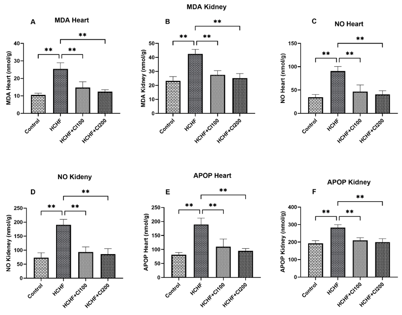
Figure 2 represents the levels of oxidative stress markers in the kidney tissues. From Figure 2A, it can be seen that the concentration of Malondialdehyde (MDA) was significantly increased in the ISO treated group when compared to the Control group (p < 0.0001). Treatment with Keora peel extract in the ISO + Keora group resulted in a significant decrease in MDA concentration when compared to the group treated with ISO only (p < 0.0001). Similarly, in Figure 2B, which represents the Nitric Oxide concentration in the kidney tissues, it was seen that the ISO treated group had a significantly higher concentration of NO compared to the Control group (p < 0.0001). Treatment with Keora peel extract in the ISO + Keora group showed a marked decrease in NO concentration when compared to the ISO administered group (p < 0.0001). Furthermore, in Figure 2C, the level of Advanced Oxidation Protein Products (AOPP) was found to be significantly increased in the ISO administered group when compared to the Control group (p < 0.0001). Keora supplementation to ISO administered rats in the ISO + Keora groups seems to have significantly decreased the AOPP concentration when compared to the ISO group (p < 0.0001). Finally, no significant difference was found between the Control group and the Control + Keora group among all Oxidative Stress markers.
Figure 2 represents the levels of oxidative stress markers in the kidney tissues. From Figure 2A, it can be seen that the concentration of Malondialdehyde (MDA) was significantly increased in the ISO treated group when compared to the Control group (p < 0.0001). Treatment with Keora peel extract in the ISO + Keora group resulted in a significant decrease in MDA concentration when compared to the group treated with ISO only (p < 0.0001). Similarly, in Figure 2B, which represents the Nitric Oxide concentration in the kidney tissues, it was seen that the ISO treated group had a significantly higher concentration of NO compared to the Control group (p < 0.0001). Treatment with Keora peel extract in the ISO + Keora group showed a marked decrease in NO concentration when compared to the ISO administered group (p < 0.0001). Furthermore, in Figure 2C, the level of Advanced Oxidation Protein Products (AOPP) was found to be significantly increased in the ISO administered group when compared to the Control group (p < 0.0001). Keora supplementation to ISO administered rats in the ISO + Keora groups seems to have significantly decreased the AOPP concentration when compared to the ISO group (p < 0.0001). Finally, no significant difference was found between the Control group and the Control + Keora group among all Oxidative Stress markers.

Figure 2: Effect of Keora peel extract on Kidney Oxidative Stress Markers: (A) MDA, (B) NO, and (C) AOPP in ISO-administered Rats. Data are expressed as mean ± Standard Error of Mean (SEM) (n=6). Statistical analysis was performed using one-way ANOVA followed by Tukey’s post hoc test. For significance, P <0.05 was selected for all measurements.
3.3. Effect of Keora Peel Extract Supplementation on Antioxidant Activity in Kidney Tissue
The antioxidant systems in the kidney tissues have been shown in Figure 3. From Figure 3A, it can be seen that the Superoxide dismutase (SOD) activity was considerably reduced in the ISO group when compared to the Control group (P = 0.0008). Conversely, the ISO + Keora group showed a significant increase in SOD activity when compared to the ISO only group (P = 0.0171). In Figure 3B, the Catalase activity of the ISO group was also significantly reduced when compared to the Control group (P = 0.0014). However, Keora peel extract treatment in the ISO administered group, ISO + Keora, showed a considerable increase in the Catalase activity levels when compared to the ISO only group (P = 0.0068). For reduced Glutathione (GSH) represented in Figure 3C, the ISO group had significantly diminished levels when compared with the Control group (P < 0.0001). ISO + Keora group exhibited a significant increase in the GSH levels when compared to the ISO only group (P = 0.0002). No statistically significant difference was found between the Control group and the Control + Keora group for any of the tested Kidney Antioxidant Activities
The antioxidant systems in the kidney tissues have been shown in Figure 3. From Figure 3A, it can be seen that the Superoxide dismutase (SOD) activity was considerably reduced in the ISO group when compared to the Control group (P = 0.0008). Conversely, the ISO + Keora group showed a significant increase in SOD activity when compared to the ISO only group (P = 0.0171). In Figure 3B, the Catalase activity of the ISO group was also significantly reduced when compared to the Control group (P = 0.0014). However, Keora peel extract treatment in the ISO administered group, ISO + Keora, showed a considerable increase in the Catalase activity levels when compared to the ISO only group (P = 0.0068). For reduced Glutathione (GSH) represented in Figure 3C, the ISO group had significantly diminished levels when compared with the Control group (P < 0.0001). ISO + Keora group exhibited a significant increase in the GSH levels when compared to the ISO only group (P = 0.0002). No statistically significant difference was found between the Control group and the Control + Keora group for any of the tested Kidney Antioxidant Activities

Figure 3: Effect of Keora peel extract on Kidney Antioxidant Activity: (A) SOD, (B) Catalase, and (C) GSH in ISO-administered Rats. Data are expressed as mean ± Standard Error of Mean (SEM) (n=6). Statistical analysis was performed using one-way ANOVA followed by Tukey’s post hoc test. For significance, P <0.05 was selected for all measurements.
3.4. Effect of Keora Peel Extract Supplementation on MPO Activity, Uric Acid, and Creatinine Levels
The results for Myeloperoxidase (MPO) activity in the kidney have been represented in Figure 4A, which shows that ISO had significantly increased the MPO activity in the kidney tissues when compared to the Control group (P < 0.0001). Treatment with Keora in the ISO + Keora group drastically reduced the elevated levels when compared to the ISO only treated group (P < 0.0001). Figure 4B representing the Uric Acid concentration in plasma also showed a significantly increased concentration of Uric Acid in the ISO group when compared with the Control group (P < 0.0001). ISO + Keora-treated group showed a significant decrease in the Uric Acid concentration when compared with the group only administered with ISO (P < 0.0001). The Creatinine Concentration in plasma, represented in Figure 4C, was highly elevated in the ISO group when compared with the Control group (P < 0.0001). The addition of Keora peel extract to ISO administered treatment group, ISO + Keora, exhibited a significantly reduced Creatinine concentration (P < 0.0001) when compared with the ISO only group. No significant difference was observed between the Control and Control + Keora groups across MPO activity, Uric Acid, and Creatinine Levels.
The results for Myeloperoxidase (MPO) activity in the kidney have been represented in Figure 4A, which shows that ISO had significantly increased the MPO activity in the kidney tissues when compared to the Control group (P < 0.0001). Treatment with Keora in the ISO + Keora group drastically reduced the elevated levels when compared to the ISO only treated group (P < 0.0001). Figure 4B representing the Uric Acid concentration in plasma also showed a significantly increased concentration of Uric Acid in the ISO group when compared with the Control group (P < 0.0001). ISO + Keora-treated group showed a significant decrease in the Uric Acid concentration when compared with the group only administered with ISO (P < 0.0001). The Creatinine Concentration in plasma, represented in Figure 4C, was highly elevated in the ISO group when compared with the Control group (P < 0.0001). The addition of Keora peel extract to ISO administered treatment group, ISO + Keora, exhibited a significantly reduced Creatinine concentration (P < 0.0001) when compared with the ISO only group. No significant difference was observed between the Control and Control + Keora groups across MPO activity, Uric Acid, and Creatinine Levels.

Figure 4: Effect of Keora peel extract on (A) MPO, (B) Uric Acid, and (C) Creatinine in ISO-administered Rats. Data are expressed as mean ± Standard Error of Mean (SEM) (n=6). Statistical analysis was performed using one-way ANOVA followed by Tukey’s post hoc test. For significance, P <0.05 was selected for all measurements.
3.4 Effect of S. apetala fruit peel extract on histology of the kidney in isoprenaline (ISO) administered rats
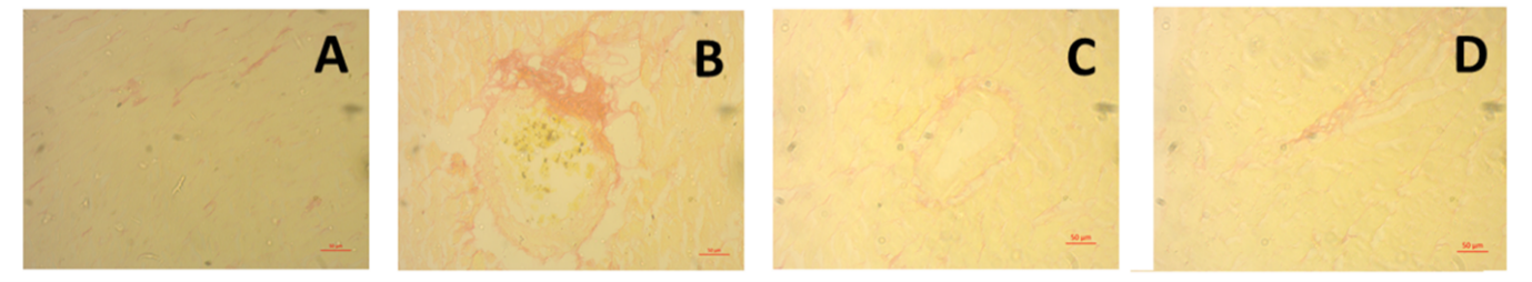
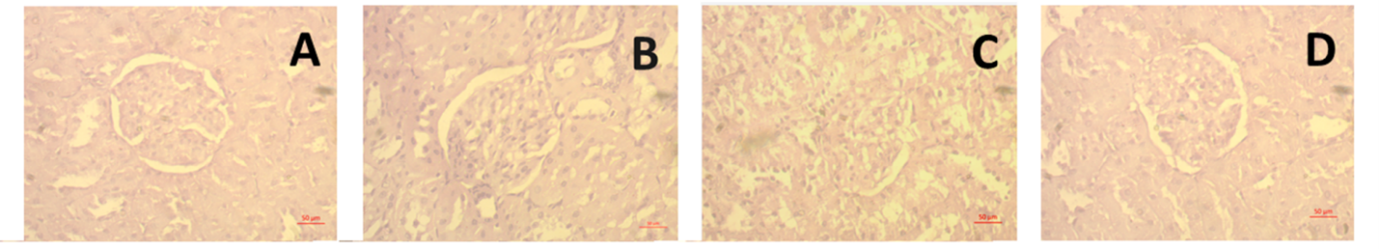
Figure 5 represents the microscopic observations from H&E and Picrosirius Red staining of kidney tissues. Hematoxylin & Eosin (H&E) staining showed that the Control (A) and Control + Keora (C) groups showed similar normal renal morphology, with the glomeruli and tubules being intact. The ISO only group (B) revealed pronounced pathological changes, including infiltration of inflammatory cells. The ISO + Keora group (D) showed preservation of the renal tissue architecture, having noticeable reduction in infiltration of inflammatory cells when compared with the ISO only group (B).
Picrosirius Red staining for collagen deposition showed minimal basal staining in the Control (E) and Control + Keora (G) groups. The ISO group (F) displayed an intense and widespread collagen deposition, mostly concentrated around the glomerulus. The ISO + Keora group (H) showed a remarkable reduction of collagen deposition when compared with the ISO only group (F).

Figure 5: Histopathological and Anti-fibrotic effect of Keora peel extract on Kidney Tissue. (A-D) H&E staining showing morphological changes (40×). (E-H) Picrosirius Red staining showing collagen deposition (20×). Groups are Control (A, E), ISO B, F), Control + Keora (C, G), and ISO + Keora (D, H)
4. Discussion
5. Conclusions
Author Contribution
Funding
Institutional Review Board Statement
Data Availability Statement
Acknowledgments
Conflicts of Interest
References
Figures