1. Introduction
2. Methodology
3. Sonneratia apetala: Mangrove Apple
4. Taxonomic Classification of S. apetala
5. Phytochemistry of S. apetala
The ability of Keora to survive and thrive in harsh, high salinity environment of mangrove forests is directly attributed to its unique and abundant profile of defensive secondary metabolites. These compounds are synthesized by the plants to combat and counteract the severe osmotic and oxidative stress given to it by its habitat [4]. Chemical screening across various plant parts consistently revealed the presence of major classes of bioactive compounds, primarily polyphenols and flavonoids, alongside significant quantities of triterpenoids, alkaloids, and tannins. The high concentration of these metabolites is the pharmacological basis for all the biological activities discussed in this review.
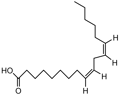
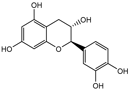
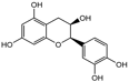
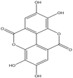
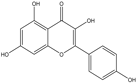
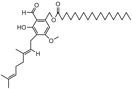
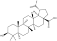
Key compounds identified through isolation and analysis include the potent polyphenols gallic acid and ellagic acid, the triterpenoids lupeol and betulinic acid, and the flavonoids quercetin and catechin. Notably, seeds and fruit pericarp are typically reported as being especially rich in polyphenols and flavonoids. A detailed, compound-by-compound breakdown of the phytochemicals isolated from S. apetala, along with their specific plant source and corresponding literature references, is presented in Tables 1 and 2.
Table 1: Chemical Constituents of the fruit of S. apetala
|
Compounds |
Activity |
Structure |
Ref. |
|
|
Phenolic compounds
|
Caffeic acid |
Antioxidant, anti-inflammatory, anticarcinogenic. |
|
[14, 15] |
|
Syringic acid |
Antioxidant, anti-inflammatory, antiproliferative, anticancer, antimicrobial, antiendotoxic. |
|
[14, 16] |
|
|
Ferulic acid |
Antioxidants, anti-inflammatory, anticarcinogenic, hepatoprotective, and antibacterial. |
|
[14, 17] |
|
|
p-Coumaric acid |
Antioxidant, anti-bacterial, antitumor, anti-inflammatory. |
|
[14, 18] |
|
|
Gallic acid |
Antioxidant, anti-inflammatory, antineoplastic. |
|
[14, 19] |
|
|
Sinapic acid |
Antioxidant, antimicrobial, anti-inflammatory, anticancer. |
|
[14, 20] |
|
|
Coumarin |
Anticoagulant, antioxidant, anti-inflammatory, antitumor, antiviral, antibacterial. |
 |
[14, 21] |
|
|
trans-Cinnamic acid |
Antioxidant, antibacterial, anti-inflammatory, antitumor. |
|
[7, 22] |
|
|
Phenol, 3,5-bis(1,1-dimethylethyl) |
Anticancer, antioxidant, antimicrobial. |
|
[7, 23] |
|
|
|
Benzenepropanoic acid |
Antioxidant, anti-inflammatory. |
|
[14] |
|
Flavonoids |
Quercetin |
Anticancer, antioxidant, anti-inflammatory, anti-cardiovascular, anti-aging, neuroprotective. |
|
[14, 24] |
|
Catechin |
Anticancer, antioxidant, anti-inflammatory. |
|
[14, 25] |
|
|
Rutin |
Antioxidant, anti-inflammatory, anti-proliferative. |
|
[14, 26] |
|
|
Myricetin |
Antioxidant, anticancer, antidiabetic and anti-inflammatory. |
|
[14, 27] |
|
|
Apigenin |
Antiproliferative, anti-inflammatory, and antimetastatic |
|
[14, 28] |
|
|
Kaempferol |
Antioxidant, anti-inflammatory, anticancer, antidiabetic, cardioprotective, neuroprotective. |
|
[7, 29] |
|
|
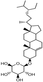
Triterpenoids |
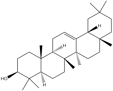
Lupeol |
Anti-inflammatory, antiprotozoal, hepatoprotective, cancer preventive.
|
|
[14, 30] |
|
|
β-amyrin |
Antioxidant. |
|
[14, 31] |
|
Others |
1,2-benzene dicarboxylic acid ester |
Anticancer, antibacterial, antidiabetic. |
|
[7] |
|
2-methyltetracosane |
Antibacterial, antioxidant. |
|
[7, 32] |
|
|
Tetratetracontane |
Antioxidant, cytoprotective, hypoglycemic, hypolipidemic, antibacterial. |
|
[7, 33] |
|
|
Heptacosane |
Antibacterial, antifungal. |
|
[7] |
|
|
2-hexyl-1-octanol |
Antimicrobial. |
|
[7] |
|
|
Vitamin |
Ascorbic acid |
Antioxidant, anti-inflammatory. |
|
[14] |
|
|
Vitamin B2 |
Antioxidant, |
|
[14] |
|
Vitamin B5 |
Antioxidant, anti-inflammatory. |
|
[14, 34] |
|
|
Vitamin B6 |
Anti-inflammatory. |
|
[14, 35] |


-png.png?width=109&height=83&name=Phenol%2c%203%2c5-bis(1%2c1-dimethylethyl)-png.png)
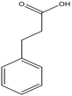


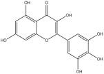


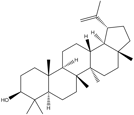
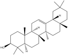


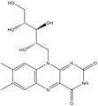
Table 2: Chemical constituents of the seed of S. apetala
|
Compounds |
Activity |
Structure |
Ref. |
|
|
Polyphenols
|
Caffeic acid |
Antioxidant, anti-inflammatory, anticarcinogenic. |
|
[6, 15] |
|
(+)-catechin |
Anticancer, antioxidant, anti-inflammatory, and anti-allergy. |
|
[6, 25] |
|
|
(-)-epicatechin |
Antioxidant, anti-inflammatory, antimicrobial, antitumor, cardioprotective. |
|
[6, 36] |
|
|
Ellagic acid |
Antioxidant, and antiadipogenic, anticancer. |
|
[6, 37] |
|
|
Gallic acid |
Antioxidant, anti-inflammatory, antineoplastic. |
|
[6, 19] |
|
|
Quercetin |
Anticancer, antioxidant, anti-inflammatory, cardioprotective, anti-aging, and neuroprotective. |
|
[6, 19] |
|
|
Rutin hydrate |
Antioxidant, anti-inflammatory, anti-proliferative. |
 |
[26, 38] |
|
|
trans-ferulic acid |
Antioxidant |
|
[38] |
|
|
trans-cinnamic acid |
Antioxidant, antibacterial, anti-inflammatory, antitumor. |
|
[22, 38] |
|
|
Myricetin |
Antioxidant, anticancer, antidiabetic, and anti-inflammatory. |
|
[38] |
|
|
Kaempferol |
Antioxidant, anti-inflammatory, anticancer, antidiabetic, cardioprotective, neuroprotective. |
|
[30, 38] |
|
|
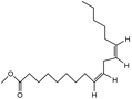
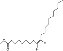
Fatty Acids |
Margaric acid |
Antitumor, antimicrobial. |
|
[39, 40] |
|
8,11-otadecadienoic acid, methyl ester |
Antimicrobial . |
|
[40, 41] |
|
|
Steric acid, methyl ester |
Antibacterial and antifungal. |
|
[40, 42] |
|
|
Linoleic acid, methyl ester |
Antioxidant. |
|
[43] |
|
|
Oleic acid, methyl ester |
Acaricidal, antimicrobial. |
|
[43, 44] |
|
|
Oleic acid |
Antifungal, antitumor. |
|
[45] |
|
|
Arachidic acid |
Anti-inflammatory, cardioprotective, anticlotting. |
 |
[40, 60] |
|
|
Linoleic acid |
Anti-inflammatory, anticoagulant, cardioprotective. |
 |
[40, 47] |
|
|
Stearic acid |
Neuroprotective, antioxidant, antilipidemic. |
[40, 48] |
||
|
Tetracosanoic acid |
Antioxidant, anticancer, antihypertensive, anti-inflammatory |
[40, 49] |
||
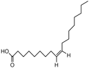
| Palmitic acid | Antioxidant, anticancer, antihypertensive, anti-infalmmatory | [12] | ||
|
Ester |
bis(2-ethylhexyl) ester |
Antimutagenic. |
|
[38, 50] |
|
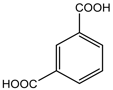
1,3-benzenedicarboxylic acid |
Catalyst. |
|
[38] |
|
|
Triterpenoid |
Lupeol |
Anti-inflammatory, anticancer, antimicrobial. |
|
[10] |
|
β-amyrin |
Antioxidant. |
|
[10, 31] |
|
|
Lupeone |
Anti-inflammatory, antiprotozoal, hepatoprotective, |
|
[10] |
|
|
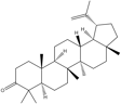
Betulinic acid |
Anti-inflammatory, antibacterial, antiviral, antidiabetic, antimalarial, antitumor |
|
[10, 52] |
|
|
Steroid |
Stigmast-5-ene 3beta |
Antidiabetic. |
|
[10, 51] |
|
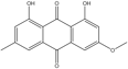
Others |
Physcion |
Anti-proliferative. |
|
[10, 53] |
|
Gibberellin |
Anti-inflammatory. |
|
[10, 54] |


%20ester-png.png?width=1789&height=455&name=bis(2-ethylhexyl)%20ester-png.png)


6. Pharmacological activities of S. apetala
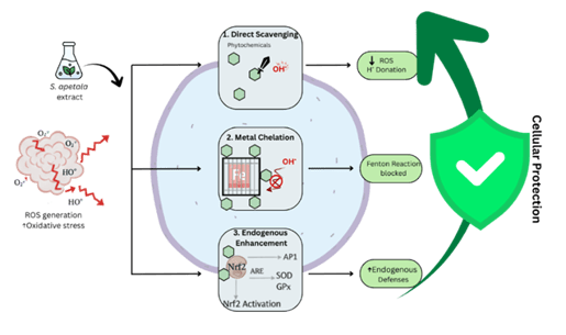
Figure 2: Generalized mechanism of plant-derived antioxidant action
The imbalance between the production of reactive oxygen species (ROS) and the body’s antioxidant defenses is termed as Oxidative stress and it is a major factor in the development of chronic disorders like diabetes, liver damage, renal dysfunction [55]. Due to the multi-targeting potential of plant-based medicines, natural antioxidants have drawn a lot of interest as possible treatment for these disorders [56]. Several studies have already evaluated the antioxidant activity of Sonneratia apetala based on both in vitro and in vivo models.
DPPH (2,2-diphenyl-1-picrylhydrazyl), ABTS (2,2’-azino-bis(3-ethylbenzothiazoline-6-sulfonic acid)), FRAP (Ferric reducing antioxidant power) and other in vitro studies have revealed a strong dose-dependent antioxidant activity in the extracts of Sonneratia apetala, thus demonstrating its free radical scavenging potential [57]. In vivo studies provided further evidence of the antioxidant activities of Sonneratia apetala extracts as administering its extracts to experimental animal models showed an increase in the activities of antioxidant enzymes like glutathione peroxidase (GPx), catalase (CAT) and Superoxide dismutase (SOD). At the same time, it lowered lipid peroxidation indicators like malondialdehyde (MDA), suggesting that the tissues were protected from oxidative damage [58].
Polyphenols and other bioactive compounds are mainly responsible for these antioxidant effects as they can usually work in three ways: they scavenge the free radicals by giving them hydrogen atoms or electrons, chelate Fe2+ ions to stop the Fenton reaction from forming hydroxyl radicals, modulation of the expression of antioxidant enzymes to reinforce cellular defense [59]. The antioxidant activities of Sonneratia apetala observed throughout various studies are summarized in Table 3, which shows the types of extracts tested, experimental models used, assays employed and key outcomes of the tests. Overall, the compiled evidences demonstrated that S. apetala showed significant antioxidant potential across multiple experimental systems, providing a strong support for its antioxidant properties.
Table 3: Antioxidant properties of Sonneratia apetala extracts
|
Model |
Treatment |
Result |
Ref. |
|
In vitro. DPPH, NO free radical scavenging assay. |
Pericarp methanolic extract. |
Showed free radical scavenging activity. |
[8] |
|
In-vitro. Reducing power and DPPH radical scavenging activity. |
Aqueous extract of fruit. |
Showed high reducing power and DPPH radical scavenging activity. |
[60] |
|
In-vitro. Colorimetric method- Fe-chelating activity. |
Fruit extract of n-hexane, chloroform, and methanol. |
All fractions showed chelating activity. Methanolic fraction showed the highest activity with IC50 of 165 μg/mL. |
[61] |
|
In-vivo. Male Swiss albino mice- 100mg/kg I.P. injection of ferric carboxymaltose induced oxidative stress. |
Methanol and n-hexane extract of fruit at 250, 500, 750, and 1000 μg/kg for 21 days. |
Reduced iron profile; highest methanolic dose completely ameliorated blood and liver iron overload and prevented oxidative stress. |
[61] |
|
In-vitro. DPPH, ABTS, and NO radical scavenging activity. |
Ethanolic fruit extract. |
Showed strong scavenging activity against ABTS, DPPH and NO radicals. |
[14] |
|
In vitro. DPPH, NO, and ABTS scavenging; In vivo. Oxidative stress mice model. |
Various fruit extracts (Pericarp methanolic, Aqueous, n-hexane, chloroform, methanol, and ethanolic). |
Extracts showed radical scavenging, reducing power, and chelating; CHCl₃ excellent (IC₅₀ 13.76 μg/mL), n-Hex & EtOAc moderate (IC₅₀ 42.03 & 49.998); highest methanol dose prevented oxidative stress in iron-overloaded mice |
[62] |
|
In vitro. DPPH free radical scavenging method. |
Methanol extract of leaf at concentrations of 5−80 μg/mL. |
strong antioxidant activity (IC50 of 41.92 μg/mL). |
[63] |
|
In vitro assays including DPPH scavenging ability, NO free radical scavenging ability, and metal chelating ability |
Methanol extracts of the fruit's seeds and pericarp |
Seed extract showed stronger antioxidant activity than pericarp, with methanolic extract being the most effective. |
[38] |
|
In vitro: DPPH, NO free radical scavenging assay. |
Pericarp methanolic extract. |
Showed free radical scavenging activity |
[64] |
|
In-vitro: Reducing power and DPPH radical scavenging activity |
Aqueous extract of fruit. |
Showed high reducing power and DPPH radical scavenging activity |
[64] |
|
In vitro: Colorimetric method - Fe-chelating activity. |
Fruit extract of n-hexane, chloroform, and methanol. |
All fractions showed chelating activity. Methanolic fraction showed the highest activity with Ic50 of 165 μg/mL |
[64] |
|
In-vivo: Male Swiss albino mice - 100mg/kg I.P. injection of ferric carboxymaltose induced oxidative stress. |
Methanol and n-hexane extract of fruit at 250, 500, 750, and 1000 μg/kg for 21 days. |
Reduced iron profile; highest methanolic dose completely ameliorated blood and liver iron overload and prevented oxidative stress |
[64] |
|
In vitro antioxidant activity using DPPH and FRAP assays |
Methanolic, ethanolic and aqueous extract of leaf |
Methanolic leaf extract showed 77.37% scavenging, ethanolic 75.14%, and aqueous 68.12% |
[65] |
|
In vitro DPPH, reducing power, and total antioxidant capacity assays |
Aqueous extract of S. apetala fruit powder |
Strong antioxidant activity: IC₅₀ = 33.5 µg/mL (DPPH), reducing power = 170.83 mg GAE/g, total capacity = 210.43 mg AAE/g. |
[66] |
|
In vivo. PO/HX-induced hyperuricemic mice. |
Aqueous extract of leaves, further concentrated with 60% ethanol |
Restored renal SOD, CAT, GSH-Px and reduced MDA and ROS in kidney tissue; ↓UA, BUN, CRE, Cys-C; |
[58] |
|
In vitro (DPPH, ABTS⁺, NO, superoxide, hydroxyl radical scavenging, Reducing power) |
Hydro-methanolic (20:80) extract of S. apetala leaves |
strong in vitro antioxidant activity, effectively scavenging |
[67] |
|
In vivo (adult male Wistar albino rats) |
Hydro-methanolic extract of Sonneratia apetala leaves |
Exhibited antioxidant effects in gastric tissue by reducing lipid peroxidation and enhancing levels of glutathione and catalase |
[67] |
|
In vitro (chemical assays: DPPH, ABTS⁺, NO, O₂⁻, HO•, Reducing power |
Aqueous extract of leaves and branches |
Showed antioxidant activity (e.g., DPPH IC₅₀ = 0.81 mg/mL, ABTS⁺ IC₅₀ = 0.16 mg/mL, others) |
[68] |
|
In vivo (Kunming mice, HUA model) |
Aqueous extract of leaves and branches (50, 100, 200 mg/kg) for 7 days |
Dose-dependent ↑ SOD, CAT, GSH; ↓ MDA, ROS in kidney; strongest effect at 200 mg/kg, surpassing BZM (CAT/GSH) and FBX (GSH); indicates potent antioxidant activity |
[68] |
|
In vitro. DPPH, total phenolic, total flavonoid, and total antioxidant capacity assays. |
Methanolic leaf extract of Sonneratia apetala |
Exhibited antioxidant activity |
[69] |
|
In vitro. DPPH, H₂O₂, hydroxyl, and superoxide radical scavenging assays; total phenolic, flavonoid, and tannin content. |
Ethanolic extract of pneumatophore (aerial root). |
Strong free radical scavenging activity (IC₅₀: DPPH 71.77 µg/ml, H₂O₂ 97.27 mg/L, OH 79.62 mg/L, O₂⁻ 108.89 mg/L |
[70] |
|
In vivo. Oral glucose tolerance test (mice). |
Ethanolic extract of pneumatophore at 250 & 500 mg/kg bw; |
Significantly reduced blood glucose at 60 and 120 min; effect comparable to standard drug |
[70] |
|
In vitro. DPPH, nitric oxide (NO) free radical scavenging assay |
Methanol fraction of seeds (MeS) |
Strong antioxidant activity from polyphenols and vitamin C. |
[12] |
|
In vitro. Lipid-soluble antioxidant assay |
n-Hexane fraction of seeds (HS) |
Antioxidant activity from lipophilic compounds (ascorbyl palmitate, fatty acids). |
[12] |
|
In vitro antioxidant assays (DPPH, free radical scavenging) |
Leaf extracts (Hexane, Ethyl acetate, Methanol) |
Methanol extract showed strongest activity; ethyl acetate moderate; hexane weak. |
[71] |
|
In vitro. Total phenolic content, DPPH free radical scavenging assay |
Crude extract, ethanol fraction, acetone fraction of S. apetala pneumatophores |
Acetone fraction showed strongest antioxidant activity (IC50 2.4 μg/mL), ethanol fraction very low, crude extract moderate.
|
[72] |
|
In vitro (DPPH, NO scavenging, reducing power, Fe²⁺ chelation, TAC) |
Methanol, diethyl ether, chloroform, and ethyl acetate fractions of Sonneratia apetala seeds |
Exhibited strong to moderate antioxidant activity; methanol seed fraction (MS) showed highest activity |
[6] |
|
In vitro: DPPH, reducing power, total antioxidant capacity (TAC), total phenolic content (TPC) |
Seeds and pericarps; fresh and stored; uncooked or cooked (2–20 min); methanol extract of seeds |
Antioxidant property: Seeds > pericarps; cooking seeds 20 min and pericarps 5–10 min gave highest activity; fresh > stored; methanol seed extract strongest |
[73] |
|
In vitro (DPPH, reducing power, Fe2+ chelation, TPC) |
Methanolic extract of bark |
methanolic bark extract showed strong antioxidant activity |
[57] |
|
In vitro antioxidant assays (DPPH, ABTS, NO scavenging, metal chelating, reducing power, total phenol, ascorbic acid, total antioxidant capacity) |
Leaf and bark extracts (acetone, ethanol, methanol, aqueous) |
Methanol leaf & bark: best DPPH, NO scavenging, reducing power; Ethanol bark: highest phenol & ascorbic acid; Ethanol leaf: highest total antioxidant capacity; metal chelating weak; overall strong antioxidant potential |
[74] |
The characteristic trait of diabetes mellitus, a group of complex metabolic disorders, is persistent hyperglycemia brought upon by either insulin resistance, a reduced production of insulin or both [75]. Ultimately, chronic hyperglycemia causes fatal problems in the liver, kidney and cardiovascular systems by inducing oxidative stress, glycation end product formation and carbohydrate metabolism dysregulation. Seeing the limitations and side effects that current synthetic antidiabetic agents can give, plant derived natural products are now in the highlights and are being increasingly investigated as an alternative or complementary therapy due to their quality to act on multiple biochemical pathways simultaneously [76].
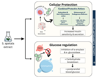
Figure 3: General antidiabetic mechanism of Sonneratia apetala extract
Sonneratia apetala has exhibited significant antidiabetic potential in both in vivo and in vitro models. In vitro models revealed that S. apetala extracts possessed potent inhibitory effects against carbohydrate digesting enzymes such as α-amylase and α-glucosidase, which delay the breakdown of carbohydrate and absorption of glucose [72, 74]. Furthermore, glucose uptake studies conducted using yeast models showed a dose dependent increase in the utilization of glucose, whereas glucose adsorption assays indicated that leaf extracts could bind to glucose molecules, consequently reducing their availability for absorption [77]. These findings together highlighted multiple mechanisms by which Sonneratia apetala can maintain or modulate blood glucose levels. In vivo studies further validated these outcomes as oral administration of the methanolic extract of fruit pericarp in streptozocin-induced diabetic rats, significantly reduced the fasting serum glucose over a prolonged treatment period [8]. Together these results suggest that both fruit and leaf derived extracts of S. apetala show hypoglycemic effects.
The antidiabetic activities of S. apetala observed across various experimental studies are summarized in Table 4, which shows the extract types, models used, and assays conducted as well as the key findings. Overall, the current evidence indicates that S. apetala holds a strong promise as a natural antidiabetic agent.
Kidney dysfunction and chronic kidney disease are often linked to underlying metabolic disorders such as diabetic nephropathy and conditions characterized by systemic oxidative stress and chronic inflammation [79]. Given that Keora possesses potent antioxidant and anti-inflammatory properties and significant anti-diabetic effects, it can be strongly positioned to have potential reno-protective agent. Even though research dedicated to its direct reno-protective effects is currently limited, the existing evidence is quite compelling [58, 68, 80].
A key study investigating the effects of the aqueous extract of S. apetala in a hyperuricemia mouse model showed robust evidence for its reno-protective potential. Hyperuricemia is one of the known causes of oxidative stress and inflammation in the kidney, often leading to kidney stone formation and renal injury. Treatment with the leaf extract showed restored antioxidant defenses (enhanced activity of antioxidant enzymes like SOD, CAT, GSH) and reduced oxidative stress markers like MDA and reactive oxygen species [58]. Similarly, seed oil extracts, branch and leaf extracts have also shown improvement in antioxidant activity, renal histology and uric acid transporters, further confirming the reno-protective potential of S. apetala [65, 77].
The general mechanism of reno-protective action is illustrated in Figure 4, and the specific experimental findings are summarized in Table 5.
Table 4 : Antidiabetic Effects of Sonneratia apetala Extracts
|
Model |
Treatment |
Result |
Ref. |
|
In-vivo. Male Long-EVANS- STZ-induced type 2 DM rats. |
Pericarp methanolic extract at 1.25g/10ml water/kg for 3 months. |
↓Reduced serum glucose level |
[8] |
|
In-vitro. α-amylase and α-glucosidase inhibition. |
10–1000 µg/mL (S. apetala fruit extract, in vitro) |
Showed strong inhibitory activity of α-amylase and α-glucosidase. |
[14] |
|
In vivo. Oral Glucose Tolerance Test (OGTT) in Swiss Albino Mice |
Methanolic extracts of 30 and 60 mg were administered; Blood samples were collected 120- and 180-min post-glucose administration. |
↓ Blood glucose levels; Significant antidiabetic potential. |
[64] |
|
In vitro. Yeast cells (Saccharomyces cerevisiae) |
Methanolic leaf extract of Sonneratia apetala (25–200 µg/mL) |
↑ Increased glucose uptake in a dose-dependent manner; highest uptake at 200 µg/mL |
[77] |
|
In vitro. α-Amylase enzyme assay |
Methanolic leaf extract of Sonneratia apetala (0.5–5 mg/mL) |
↓ Inhibited α-amylase; near 100% inhibition at 2 mg/mL |
[77] |
|
In vitro. Glucose adsorption assay |
Methanolic leaf extract of Sonneratia apetala (1%) |
↑ Adsorbed glucose proportionally to concentration; maximum at 100 mmol/L |
[77] |
|
In vivo. Mice (oral glucose tolerance test) |
Ethanolic extract of pneumatophores of Sonneratia apetala (250 & 500 mg/kg) |
↓ Blood glucose significantly at 60 and 120 min; |
[70] |
|
In vitro. α-Glucosidase inhibition |
Chloroform: Methanol (1:1) extract of leaves |
↓ Inhibited α-glucosidase activity (IC50 = 286 µg/mL) |
[78] |
|
In-vitro. α-amylase inhibition & Raphanus sativus root-growth inhibition |
Pneumatophore crude methanol extract, ethanol fraction (95%), acetone fraction (50% acetone) |
Crude: moderate α-amylase & root inhibition; Ethanol: low; Acetone: strong α-amylase & root inhibition (tannin-rich). |
[72] |
|
In vitro, Yeast α-glucosidase assay |
Leaf & bark extracts: Acetone, Ethanol, Methanol, Aqueous |
↓ Inhibited α-glucosidase activity in a dose-dependent manner; methanol extract most potent |
[74] |
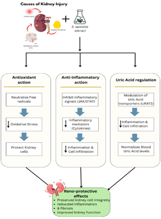
Figure 4: Flowchart illustrating the general reno-protective mechanism of S. apetala extracts
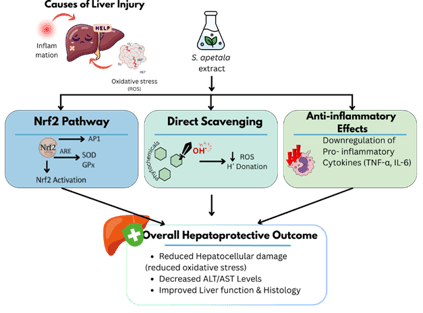
Figure 5: General Hepatoprotective mechanism of S. apetala Extract
Table 5 : Reno-protective effects of Sonneratia apetala extracts
|
Model |
|
Treatment |
|
Result |
|
Ref. |
|
In vivo. PO/HX-induced hyperuricemic mice. |
|
Aqueous extract of leaves, further concentrated with 60% ethanol |
|
Restored renal SOD, CAT, GSH-Px and reduced MDA and ROS in kidney tissue; ↓UA, BUN, CRE, Cys-C; |
|
[58] |
|
In vivo. Potassium oxonate/hypoxanthine-induced hyperuricemic mice |
|
Sonneratia apetala seed oil (SSO) |
|
↓ Serum UA, CRE, and BUN. ↑ SOD, CAT, GSH-Px with ↓ ROS and MDA levels. ↓ Kidney histopathological lesions. ↓ MCP-1, IL-1B, IL-6, IL-18, TNF-a. |
|
[80] |
|
In vivo. Hyperuricemia mice induced by Potassium oxonate (PO) and hypoxanthine (HX) |
|
Aqueous extract of leaves and branches |
|
↓ Kidney weight and index. ↓ Serum UA, CRE, and BUN. ↓ Kidney histopathological changes. ↓ MDA and ↑ CAT, SOD, and GSH. ↓ Renal inflammatory markers (IL-6, IL-18, IL-1ẞ, TNF-α, MCP-1, TGF-β). Regulated renal uric acid transporters (OAT1, URAT1, GLUT9). |
|
[68] |
|
In vivo. Isoprenaline-induced male Long-Evans rats |
|
Keora fruit peel extract 100 mg/kg/day |
|
↓MDA, NO, and AOPP levels in the kidney ↑CAT, SOD, and GSH activity in the kidney ↓MPO activity in the kidney ↓Creatinine and uric acid levels |
|
[81] |
Table 6: Hepatoprotective effects of Sonneratia apetala extracts
|
Model |
|
Treatment |
|
Result |
|
Ref. |
|
In-vivo. Male Kunming mice- acetaminophen-induced liver injury. |
|
Aqueous fruit extract at 100, 200, and 400mg/kg/day orally for 1 week. |
|
↑ Survival, ameliorated liver histology, ↓ ALT, AST, MDA, TNF-α, IL-6, MPO; ↑ GSH, GSH-Px, CAT, total antioxidant capacity |
|
[60] |
|
In vivo: Male Swiss albino mice with iron overload induced by ferric carboxymaltose |
|
In vivo: S. apetala fruit extract fractions (Hex, Chl, Met) at 100, 500, and 1,000 µg/kg bw daily for 15 days |
|
All fractions (Hex, Chl, Met) showed dose-dependent amelioration of iron overload. Met was most effective, completely ameliorating iron overload |
|
[61] |
|
In vivo (Kunming mice, HUA model) |
|
Aqueous extract of leaves and branches (50, 100, 200 mg/kg) for 7 days |
|
↓ UA, CRE, and BUN in serum. |
|
[68] |
|
In vivo. PO/HX-induced hyperuricemic mice. |
|
Aqueous extract of leaves, further concentrated with 60% ethanol |
|
No hepatotoxicity; ↓ hepatic XOD and ADA activities |
|
[58] |
Liver playing a crucial role in the metabolism and detoxification of substances like alcohol, drugs and metabolic byproducts makes it highly susceptible to injury and diseases, leading to conditions like NAFLD, fibrosis, or other metabolic disorders [82]. Hepatotoxicity is commonly associated with an elevated level of liver enzymes such as alanine aminotransferase (ALT) and aspartate aminotransferase (AST), lipid peroxidation, and inflammatory responses. Although synthetic hepatoprotective medications are available, prolonged use of such medicines may result in adverse side effects, which has led our researchers to the exploration of plant-derived natural products as alternatives or supplementary treatments [60, 61].
Sonneratia apetala has demonstrated significant hepatoprotective potential in various experimental models. In vivo studies using male Kunming mice with acetaminophen induced liver injury showed that the oral administration of the aqueous extract of fruit at 100, 200 and 400 mg/kg/day for 1 week significantly increased survival rats, ameliorated histological liver damage and also reduced the elevated levels of ALT, AST, malondialdehyde (MDA), TNF-α, IL-6 and myeloperoxidase (MPO). Alternatively, antioxidant markers including Glutathione (GSH), glutathione peroxidase (GSH-Px), catalase (CAT) and total antioxidant capacity were significantly increased, indicating a strong antioxidant and anti-inflammatory effects in the liver [60]. In addition to modulating oxidative stress, S. apetala extracts have also shown influence in purine metabolism by inhibiting hepatic xanthine oxidase (XOD) and adenosine deaminase (ADA), ultimately reducing the uric acid production and improving the renal-hepatic function in hyperuricemic mice models [58, 68]. Collectively, these findings portray the various ways S. apetala extracts can exert their protective effects on the liver. Figure 5 provides a comprehensive overview of S. apetala’s hepatoprotective effects. A comprehensive overview of different in vivo studies illustrating the hepatoprotective potential of S. apetala is provided in Table 6.