Original article Open access | J. Bio. Exp. Pharm. 2026, 4(1), 42-57 | https://doi.org/10.62624/JBEP00.0033
Investigation of the biological activities of synthetic dihydropyridine analogues of felodipine: An in vitro analysis
Juhaer Anjum1, Rumman Reza1, Safaet Alam1, Md. Saiful Islam2, Shaila Kabir1, Mohammad Rashedul Haque1*

Abstract

The most urgent medical concerns worldwide are hypertension and the cardiovascular disorders it causes. Many drugs have been created and introduced to the market to treat hypertension. In most countries, hypertension is a significant health concern because of its impact on patient mortality and morbidity. Since calcium channel blockers have been demonstrated to not increase the risk of coronary events and stroke, they appear to be a good choice for treating hypertension both alone and in conjunction with other agent classes. They might have additional benefits besides decreasing blood pressure. Drugs with a dihydropyridine nucleus in their chemical structures are known as dihydropyridine calcium channel blocker. The Hantzsch reaction is necessary for the synthesis of 1,4-dihydropyridines, which are used to treat vascular issues, cancer, and hypertension. Other analogues can be created by altering the ester groups at the 3 and 5 positions of the dihydropyridine core or the ortho/meta/para substitutions on the benzaldehyde ring (for instance, substituting the dichlorophenyl group of felodipine to other substituted phenyl rings). These analogues have recently been synthesized using p-toluenesulfonic acid as a catalyst in solvent-free conditions, producing good yields (80–96%) in short reaction times. Four analogues of felodipine were created for the current study, and the compounds' biological activities were examined. Evaluating the biological potential of the chemicals presented was the primary goal of this investigation. The efficacy of the synthesized compounds was assessed using biological activity assays, including DPPH assay, thrombolytic assay, alpha-amylase inhibitory assay, antimicrobial testing by disc diffusion method, and cytotoxic analysis.


Keywords

Anti-diarrheal activity
castor oil induced diarrhea
Hypertension
cardiovascular disease
calcium channel blocker
dihydropyridine nucleus
The Hantzsch reaction

    1. Introduction

    Globally, hypertension and its associated cardiovascular problems continue to be the most pressing medical issues [1]. To treat hypertension, a number of medications have been developed and put on the market [2]. Due to its effects on patient mortality and morbidity, hypertension is still a major health issue in the majority of nations. In fact, hypertension is responsible for 1.4% of disability-adjusted life years, 1.9% of years of life lost, and nearly 5.8% of all deaths worldwide [3]. As a result, preventing and controlling hypertension in the community is today a critical issue. A thiazide-type diuretic, calcium  (Ca2+)  channel blocker (CCB), angiotensin-converting enzyme inhibitor (ACEI), or angiotensin receptor blocker (ARB) should be considered as an initial antihypertensive medication in the non-black population, in addition to the guidelines that pharmacologic treatment should be started when blood pressure (BP) is 150/90 mmHg or higher in adults under 60 years of age, or 140/90 mmHg or higher in patients with hypertension and diabetes [4]. One of the most crucial starting medications for antihypertensive monotherapy is CCB [5].

    CCBs seem to be a good option for both monotherapy and combination with other agent classes in the treatment of hypertension since they have been shown to not raise the risk of coronary events and stroke [6]. They may also offer particular advantages beyond lowering blood pressure. In China and other Eastern Asian nations, dihydropyridine (DHP) CCBs are currently among the most often prescribed antihypertensive drugs [7]. Dihydropyridine CCBs are the medications that contain dihydropyridine nucleus in their chemical structures [8]. These medications are responsible for reducing blood pressure by relaxing blood vessels via calcium entry blockade [9]. These are primarily used as first line therapy for high blood pressure ((hypertension) and angina whereby, these work by reducing resistance to blood flow through vasodilation. Some examples of dihydropyridine ring containing calcium channel blockers include amlodipine, nifedipine, nisoldipine, felodipine and nimodipine [10]. In order to control hypertension, several of these medications have a dihydropyridine skeleton structure, function as effective calcium channel blockers, and alter the intake of calcium ions in vascular smooth muscle [11]. The synthesis of such moieties is essential, and it is currently a focus to describe their structure–activity relationship, their evolved and sophisticated synthetic processes, and future potential in this field. SAR analogy has been essential in creating compounds with greater potency because the classic SAR is a helpful tool in the hunt for new medications [12].

    Felodipine belongs to dihydropyridine class of calcium channel blockers [10]. It is useful in managing essential hypertension and also can be used to reduce the risk of cardiovascular mortality and morbidity. One of the major benefits of felodipine usage is the impact on reducing the occurrence of strokes [13]. In cases of patients with mild-to-moderate hypertension, controlled release monotherapy of felodipine shows efficacy compared to beta blockers, angiotensinogen converting enzyme (ACE) inhibitors or other calcium channel blockers. Patients suffering from severe hypertension who are unresponsive to beta blockers and diuretics, felodipine controlled release can be considered as add-on therapy [14]. Felodipine is a dihydropyridine derivative, more precisely the ethyl methyl ester of 4-(2,3-dichlorophenyl)-1,4-dihydro-2,6-dimethyl-3,5-pyridinedicarboxylic acid. Its molecular weight is 384.26 and its formula is C18H19Cl2NO4. It has two methyl groups, two ester groups (ethyl and methyl), a core dihydropyridine ring, and a 2,3-dichlorophenyl group that blocks calcium channels and is bonded at the 4-position. Important structural characteristics include the basic heterocyclic ring structure, the dihydropyridine ring. Methyl groups at positions two and six, as well as ester groups (ethyl and methyl) at positions three and five, are substituents. A 2,3-dichlorophenyl group is located at 4-position.

     Nitrendipine, lercanidipine, and more recent derivatives like elnadipine are examples of felodipine analogues, which are related dihydropyridine calcium channel blockers (CCBs) that share the core structure of 3,5-pyridinedicarboxylic acid esters but differ in substituents (such as the dichlorophenyl group or ester groups) to alter their potency, selectivity, and duration of action [15]. They all have a similar core ring structure with particular ester groups and a substituted phenyl ring at the 4-position since they are all 1,4-dihydropyridine derivatives. Similar to felodipine, they reduce blood pressure by relaxing and widening blood arteries by blocking L-type calcium channels in vascular smooth muscle [15]. The main goal is to manage essential hypertension, or elevated blood pressure.

    In the present study, four dihydropyridine analogues were synthesized using Hantzsch reaction. The classic process for creating 1,4-dihydropyridine calcium channel blockers is the Hantzsch dihydropyridine synthesis, also known as the Hantzsch reaction or condensation, which is commonly used to create felodipine and its derivatives. An aromatic aldehyde, a beta-keto ester (like methyl or ethyl acetoacetate), and a nitrogen supply (often ammonium acetate or ammonia) are typically involved in this multicomponent reaction (MCR). Specifically, felodipine is made by condensing 2,3-dichlorobenzaldehyde, methyl acetoacetate, and ethyl acetoacetate with ammonia, usually in an alcoholic solvent. To create the dihydropyridine ring, the process first undergoes a Knoevenagel condensation, then a Michael addition and cyclisation.

    The main aim of the current study is to find biologically active molecules that can be developed as drugs in future. Four dihydropyridine analogues were synthesized using Hantzsch reaction. Biological activity tests such as DPPH assay, thrombolytic assay, alpha-amylase inhibitory assay, antimicrobial assay by disc diffusion method and cytotoxic analysis were carried out to evaluated the effectiveness of the synthesized compounds.

    2. Materials and Methods

    2.1 DPPH assay

    2.0 mL of the methanolic solution of the test substances at different concentrations (500 μg/mL to 5 μg/mL) were mixed with 3.0 mL of the prepared DPPH methanol solution of 20 μg/mL concentration. The reaction was left to conduct for 30 minutes at room temperature in a dark, light-proof chamber. Absorbance values were taken after the aforementioned time period at 517 nm wavelength against methanol using a UV spectrophotometer.

    Inhibition of free radical DPPH in percent (I%) was calculated using the following formula:

                                                                                                                                 (I%) = (1 – Asample/Ablank) X 100

    Here, Ablank is the absorbance of the control reaction that contains all reagents except the test material. Test substance concentration providing 50% inhibition (IC50) was calculated from the graph where inhibition percentage was plotted against test substance concentration.

    2.2 α-Amylase Inhibitory Assay

    α-Amylase inhibitory activity can be assayed through a colorimetric reaction employing the reagent 3,5-dinitrosalicylic acid (DNS). The experiment colorimetrically quantitates maltose released from starch through the reduction of DNS. In the presence of substances that inhibit the α-amylase enzyme, maltose production is reduced, and this reduction is reflected in the colorimetric assay.

    Maltose reduces the alkaline 3,5-Dinitro salicylic acid (DNS) and changes its pale-yellow color to orange-red. The intensity of this orange-red color is concentration-dependent and therefore allows the quantization of maltose in the sample. In the presence of α-amylase and starch, the quantity of maltose is directly proportional to the enzymatic activity, allowing for a rapid and accurate method to assess α-amylase enzymatic activity.

    2.3 Anti-microbial assay by disk diffusion method

    The antimicrobial screening is performed to ascertain the susceptibility of various bacteria to any agent of interest. This test is considered to be the first stage of antimicrobial drug research. This test measures the ability of a test sample to inhibit in vitro bacterial growth in a control culture medium. The four test substances were dissolved in ultrapure water to form 1500µg/mL solutions. These solutions were loaded into the diffusion disks for the assay. One species of gram-negative bacteria, Escherichia coli, and one species of gram-positive bacteria, Staphylococcus aureus, were employed for the disc diffusion assay. The Mueller-Hinton agar media (ThermoFisher Scientific) was used for the bacterial cultures [16].

    2.4 Cytotoxic Activity

    Cytotoxic effect was examined in the Centre for Advanced Research in Sciences (CARS), University of Dhaka, using their commercial services. In brief, Vero, a kidney epithelial cell line extracted from an African green monkey, was maintained in DMEM (Dulbecco's Modified Eagles' medium) containing 1% penicillin- streptomycin (1:1) and 0.2% gentamycin and 10% fetal bovine serum (FBS). Cells (3.0×10⁴/200µl) were seeded onto a 48-well plate and incubated at 37°C + 5% CO2. The next day, 50 µl sample (filtered) was added to each well. Cytotoxicity was examined under an inverted light microscope after 48h of incubation. Duplicate wells were used for each sample.

    2.5 Thrombolytic activity

    100μL of the test substances, the control (streptokinase), and blank (sterile, ultrapure water) were added to tubes holding serum-free blood clots of known weight. All of the tubes were incubated for 90 minutes at 37°C to facilitate clot lysis [17]. After incubation, the fluid that had leaked out of the clots was collected without disturbing the clots, and the tubes were weighed again to assess the weight difference after the clot had been disrupted by the administration of the test substances. The difference in weights between those obtained before and after clot lysis was expressed as a percentage of clot lysis, as shown below:

    Percentage of clot lysis = (Weight of the clot after lysis / Weight of clot before lysis) × 100

    3. Results

    3.1 Chemical structure of dihydropyridine analogues

    The class of calcium channel blockers known as 1,4-Dihydropyridines (DHPs) is distinguished by a core, nitrogen-containing, semi-unsaturated six-membered ring that serves as a "privileged scaffold" for cardiovascular medications such as amlodipine and nifedipine. They mainly affect L-type voltage-gated calcium channels and are characterized by a substituted 4-aryl-1,4-dihydropyridine-3,5-dicarboxylate structure. Occasionally referred to as C5H7N or derivatives, six-membered 1,4-Dihydropyridine ring has a nitrogen atom (NH) at position 1 and two double bonds at positions 2 and 3, as well as 5 and 6. In most cases, the 1,4-DHP ring takes on a flattened-boat shape. Lower alkyl groups typically methyl, or -CH3 occupy substituents at positions two and six, which are essential for activity. An important characteristic is a phenyl ring at the 4-position, which is usually replaced at the ortho or meta locations by electron-withdrawing groups (such as nitro, -NO2, or chloro, -Cl). To maximize calcium channel blocking activity, the 3- and 5-positions need carboxylate groups, which are usually esterified. Tissue selectivity and potency are affected by substituents on the 4-Aryl ring, especially at the ortho or meta locations. One crucial structural factor for binding is the NH group at the 1-position. Its calcium-blocking action is eliminated when the ring oxidizes to a pyridine (losing the NH). The 4-carbon becomes chiral when the esters at the 3- and 5-positions differ. As a blocker, the (S)-enantiomer frequently shows more activity. The structures of synthesized compounds are shown in Figure 1.
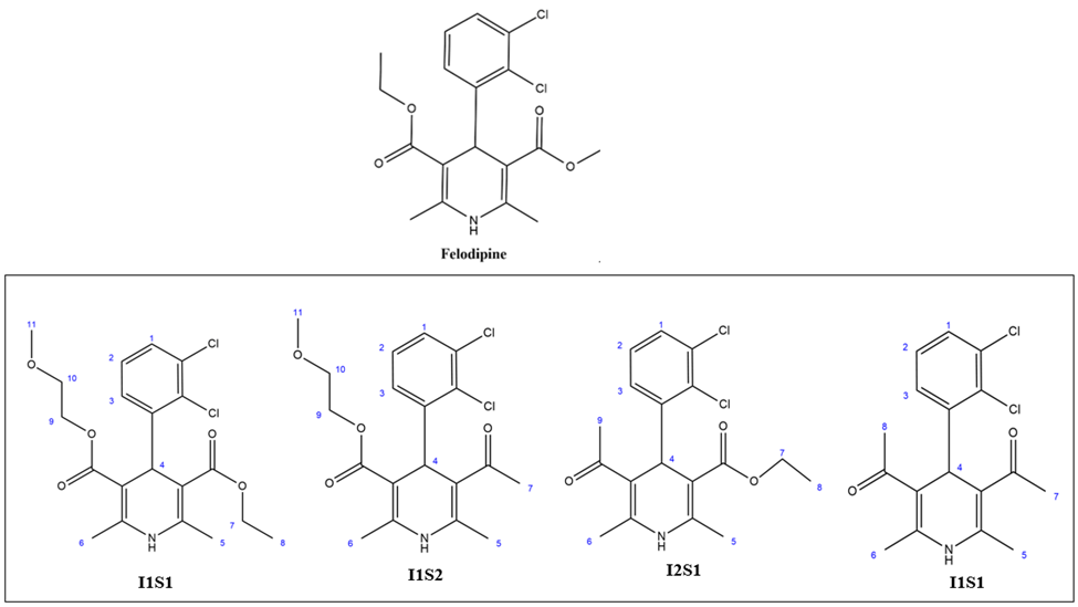
    Figure 1. Chemical structures of dihydropyridine analogues.

    1.  

    3.2 DPPH radical scavenging assay of dihydropyridine analogues

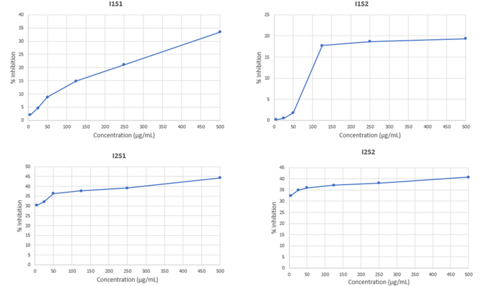
    In the DPPH assay, the control ASA displayed an IC50 value of 31.33μg/mL. However, none of the synthesized test molecules fared well in the test. Compound I2S1 displayed the most promising results in the group, displaying 44.31% inhibition at the maximum concentration of 500μg/mL as shown in Figure 3. Compound I2S2 followed suit, displaying 40.74% inhibition at the maximum concentration. Compound I1S1 displayed a 33.44% inhibition at the maximum concentration, and compound I2S2 displayed a 19.31% inhibition at the maximum concentration as shown in Table 1. The compounds were therefore not found to be promising in terms of antioxidant activity based on the assay results. Compounds of this category have, however, been previously reported to possess significant antioxidant properties. Sugawara et al. experimented on felodipine and its analogs benidipine, nisoldipine, nilvadipine, nicardipine, nifedipine, and nitrendipine; exploring their capacity to provide antioxidant effects on rat myocardial membrane lipid peroxidation, and found that nilvadipine, nisoldipine, felodipine, nicardipine, and benidipine was promising in terms of antioxidant activity [15]. The synthesized test molecules in question may therefore contain some antioxidant activity, however, further experimentation will be required in this direction to reach a concrete decision.
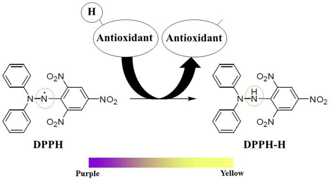
    Figure 2. Principle of the DPPH antioxidant assay.

    Table 1. IC50 value determination of standard and test substances 

    Group

    Concentration (μg/mL)

    Absorbance

    % Inhibition

    IC50 (μg/mL)

    Blank

    ASA

    500

    0.041

    93.34416

    31.33

    0.616

    250

    0.062

    89.93506

    125

    0.076

    87.66234

    50

    0.171

    72.24026

    25

    0.252

    59.09091

    5

    0.423

    31.33117


    I1S1

    500

    0.41

    33.44156

    N/A

    0.616

    250

    0.486

    21.1039

    125

    0.524

    14.93506

    50

    0.562

    8.766234

    25

    0.587

    4.707792

    5

    0.603

    2.11039


    I1S2

    500

    0.497

    19.31818

    N/A

    0.616

    250

    0.501

    18.66883

    125

    0.507

    17.69481

    50

    0.605

    1.785714

    25

    0.613

    0.487013

    5

    0.615

    0.162338


    I2S1

    500

    0.343

    44.31818

    N/A

    0.616

    250

    0.375

    39.12338

    125

    0.384

    37.66234

    50

    0.392

    36.36364

    25

    0.418

    32.14286

    5

    0.429

    30.35714


    I2S2

    500

    0.365

    40.74675

    N/A

    0.616

    250

    0.381

    38.14935

    125

    0.387

    37.17532

    50

    0.394

    36.03896

    25

    0.401

    34.9026

    5

    0.416

    32.46753

    Figure 3: Percent inhibition for different concentrations of compounds I1S1, I1S2, I2S1 and I2S2 from DPPH assay.

    3.3 Evaluation of Antidiabetic Activity: α-Amylase Inhibitory Assay

    In this assay, compound I2S1 and I1S2 performed reasonably well, the first displaying a 49.68% inhibition of α-amylase and the second 33.54% at the maximum concentration. This inhibition was observed to be dose dependent. The data from the α-amylase inhibition assay has been presented in Figure 5.
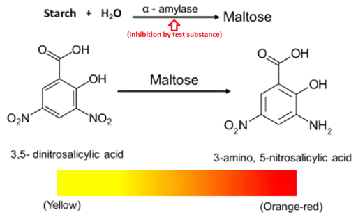
     Figure 4. Principle of the α-amylase inhibitory assay.
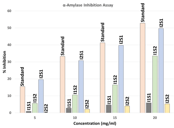
    Figure 5. Percentage of α-Amylase inhibition by standard and test substances.

    3.4 Evaluation of Antimicrobial Activity: Disc Diffusion Method

    The antimicrobial screening is performed to ascertain the susceptibility of various bacteria to any agent of interest. This test is considered to be the first stage of antimicrobial drug research. This test measures the ability of a test sample to inhibit in vitro bacterial growth in a control culture medium. Despite the vast usefulness and widespread application of the test, the test offers little ground for universal standardization. In practical research, the diameter of the zone of inhibition or the minimum quantity of a test substance required to inhibit bacterial growth has been used to report antibacterial activity in a quantitative fashion. These parameters, however, are subject to wide fluctuations depending on confounding variables such as inoculum volume, culture medium composition, pH, and incubation temperature [18]. The antimicrobial assay can be executed by any of the following three methods: disc diffusion method, serial dilution method, or bio-autographic method. Among the above-mentioned techniques, the disc diffusion method is widely accepted for preliminary screening of test agents that may possess antimicrobial activity. It is essentially a quantitative or qualitative test indicating the sensitivity or resistance of the microorganisms to the test materials. This method, however, does not offer any distinction between bacteriostatic and bactericidal activity [19].

    The four test substances were dissolved in ultrapure water to form 1500 µg/mL solutions. These solutions were loaded into the diffusion disks for the assay. One species of Gram-negative bacteria, Escherichia coli, and one species of Gram-positive bacteria, Staphylococcus aureus, were employed for the disc diffusion assay. The Mueller-Hinton agar medium (Thermo Fisher Scientific) was used for the bacterial cultures. The media formulation is provided in Table 2.

    Table 2. Composition of the Mueller-Hinton agar medium

    Formulation Component

    Concentration (gm/L)

    Dehydrated infusion of beef

    300

    Casein hydrolysate

    17.5

    Starch

    1.5

    Agar

    17

    pH 7.3 ± 0.1

    Temperature 25°C


    38 g of the ready-mix medium was added to 1 L of ultrapure water. The medium was sterilized through autoclaving at 121°C for 15 minutes. All procedures were carried out in a laminar airflow hood to minimize chances of contamination. The inner surface of the hood was disinfected with ethanol, and the inbuilt UV lamp was also used to the same end. Petri dishes and other associated glassware were disinfected at 121°C for 15 minutes. UV lamp sterilization was also carried out on the micropipette tips, cotton, forceps, blank discs, and any other associated apparatus that was not fit for autoclaving. Sub-cultures of the bacteria of interest were prepared from the pure cultures through inoculation under a sterile laminar air hood with the use of a sterile inoculation transfer loop. The inoculated agar slants were incubated for 24 hours at 37°C for optimum growth. These fresh cultures were used for the sensitivity test. The test organisms were transferred from the subculture to the test tubes containing about 10 mL of melted and sterilized agar medium with the help of a sterilized transfer loop in an aseptic area. The test tubes were shaken by rotation to get a uniform suspension of the organisms. The bacterial and fungal suspension was immediately transferred to the sterilized Petri dishes. The Petri dishes were rotated several times clockwise and anticlockwise to ensure a homogeneous distribution of the test organisms in the media [20].

    Sterilized metrical (BBL, Cocksville, USA) filter paper discs were taken in a blank petri dish under the laminar hood. Then, discs were soaked with 20 µL of the test sample solutions and dried, resulting in the discs being loaded with 30 µg of the test samples each. Standard Chloramphenicol (30 μg/disc) discs were used as the positive control to compare the activity of a standard antibiotic against the test organisms to that exerted by the test substances. Blank discs were used as negative controls to ensure that the residual solvents (left over in the discs even after air-drying) and the filter paper were not active themselves. After application of the discs, the Petri dishes were incubated in a sterile environment at 37°C for 24 hours [18]. After the incubation period, the diameter of the zone of inhibition was measured using a slide caliper to ensure accuracy.

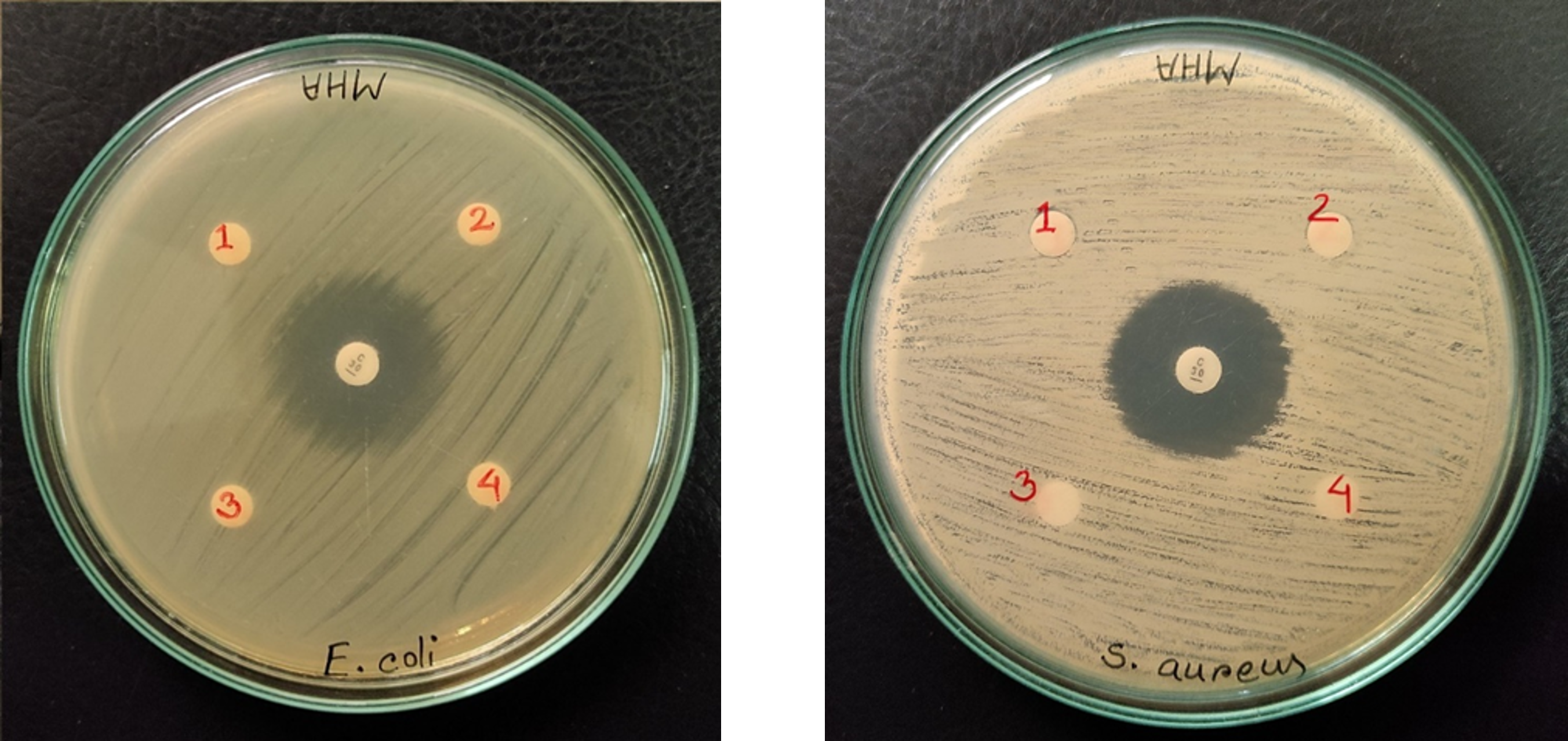
    The four synthesized test substances displayed no antimicrobial activity in either the Gram-positive Staphylococcus aureus strain or the Gram-negative Escherichia coli strain. These findings were found to be parallel to previous reports. Although some dihydropyridine analogs have been reported for their antimicrobial activity, these analogs are mostly sulfur-containing compounds or compounds with aromatic side rings in the dihydropyridine ring, whereas our synthesized analogs are structurally dissimilar [21,22]. Venkataraman et al. reported their synthesized dihydropyridine analogs PNN and PAH to possess antimicrobial properties against Staphylococcus aureus and Escherichia coli, respectively [22]. These compounds, however, share little structural similarity with our synthesized compounds. The antimicrobial assay data have been provided in Table 3.
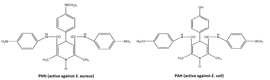
     

    Figure 6. Chemical Structures of compounds PNN and PAH [22].

    Table 3. Antimicrobial activity of the test samples

    Test Organisms

    Diameter of the Zone of Inhibition (mm)

    I1S1

    I1S2

    I2S1

    I2S2

    Chloramphenicol

    E. coli (gram-negative)

    -

    -

    -

    -

    26

    S. aureus (gram-positive)

    -

    -

    -

    -

    24


    Figure 7. Zone of inhibition of the test substances: A) E. coli and B) S. aureus

    3.5 Assessment of Cytotoxic Activity

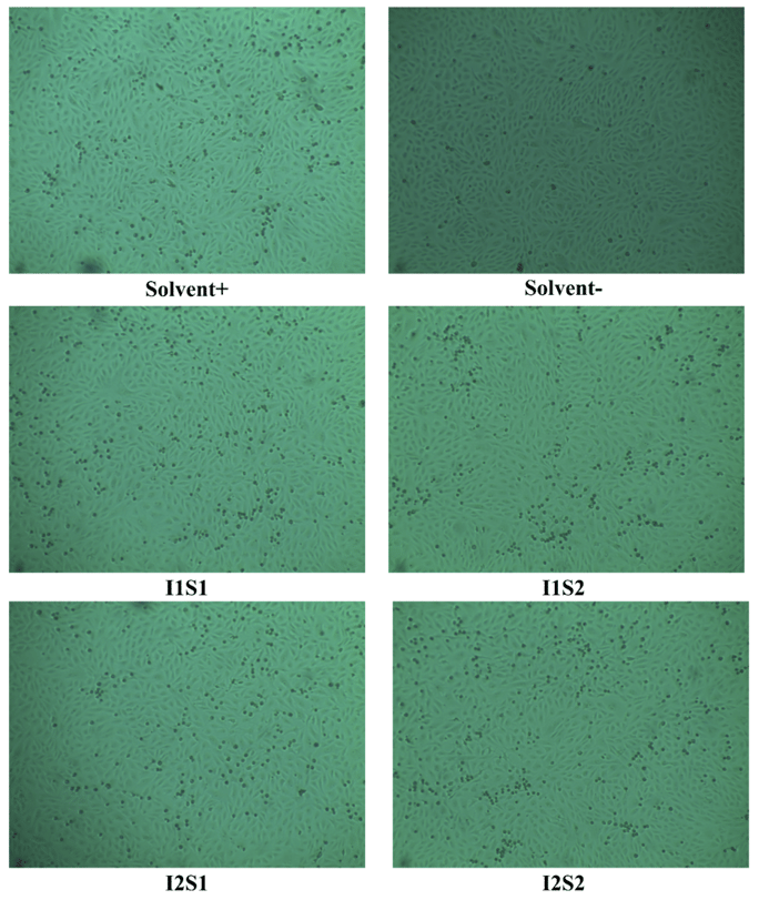
    The synthesized drug analogs did not produce any cytotoxicity on the Vero cell line. Cell survival rates were greater than 95% for all samples as shown in Figure 8. Felodipine itself has also been reported to not be cytotoxic in multiple high-throughput screening assays (PubChem Bioassay Record for AID 1825; PubChem Bioassay Record for AID 1345086). The data from the cytotoxicity assay has been presented in Table 4.

     Table 4. Cytotoxicity assay of standard and test substances 

    Sample code

    Survival of Vero Cells

    Remarks

    Solvent -

    100%

    No cytotoxicity was observed on the Vero cell line in any of the test samples.

    Solvent +

    >95%

    I1S1

    >95%

    I1S2

    >95%

    I2S1

    >95%

    I2S2

    >95%

    Figure 8 Cytotoxicity assay of the test samples using Vero cells.

    3.5 Thrombolytic assay

    Thrombus (blood clot) formation in the circulatory system can result in athero-thrombotic diseases such as acute cerebral or myocardial infarction. Such complications are often associated with other prevalent conditions such as obesity, hypertension, hyperlipidemia, hypercholesterolemia, and atherosclerosis; and can lead to morbidity or mortality [23]. Bioactive molecules that provide thrombolytic effects are therefore in high demand. To assess the thrombolytic activity of the test substances, the test substances were applied to a thrombus (blood clot) of known weight, and after a certain period of rest, the amount of clot lysis was observed through weighing. The data from the test substances were compared to that of the control streptokinase [17].

    Test substances were dissolved in ultrapure water to form 100μg/mL solutions. 1,500,000 I.U. of commercially available lyophilized alteplase (streptokinase) was collected, and resuspended by mixing thoroughly with 5 ml of sterile ultrapure water. From this suspension, 100μL of suspension containing 30,000 I.U. of the enzyme was used for the thrombolysis assay. 10 mL of whole veinous blood was collected from a healthy human volunteer, and 6 previously weighed Eppendorf (Ep.) tubes were filled with the collected blood, each having 1 mL of blood. The tubes were incubated at 37°C for 45 minutes, and the serum was removed completely without disturbing the clot. The weight of the clots was determined by reweighing the clot-filled Eppendorf tubes and subtracting the weight of the empty Eppendorf tubes from this. The following formula illustrates this mathematical operation:

    Clot weight = Weight of clot containing tube - Weight of tube alone

    100μL of the test substances, the control (streptokinase), and blank (sterile, ultrapure water) were added to tubes holding serum-free blood clots of known weight. All of the tubes were incubated for 90 minutes at 37°C to facilitate clot lysis. After incubation, the fluid that had leaked out of the clots was collected without disturbing the clots, and the tubes were weighed again to assess the weight difference after the clot had been disrupted by the administration of the test substances. The difference in weights between those obtained before and after clot lysis was expressed as a percentage of clot lysis, as shown below:

    Percent of clot lysis = (Weight of the clot after lysis / Weight of clot before lysis) × 100

    In the thrombolytic assay, compounds I2S2, I1S2, and I2S1 displayed mild thrombolytic effect; the first showing a 20.86% clot lysis, the second a 14.53% lysis, and the third a 11.76% lysis. Compound I1S1 displayed negligible thrombolytic activity at 2.21% lysis as shown in figure 9. Our findings were found to corroborate previous literature. Di Vergouwen et al. reported that dihydropyridine calcium-channel antagonists (structural analogs of our synthesized compounds) such as felodipine, nimodipine, amlodipine, and cilnidipine significantly stimulate endogenous fibrinolytic activity. However, this fibrinolytic activity was reported to be considerably weaker compared to the activity exerted by fibrinolytic drugs such as recombinant tissue plasminogen activator and urokinase. 
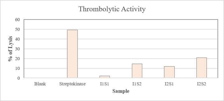
     Figure 9. Thrombolytic activity of the test samples. 


    4. Discussion

    The production of 1,4-dihydropyridines, which are used to treat hypertension, cancer, and vascular problems, depends on the Hantzsch reaction [24]. By changing the ortho/meta/para substitutions on the benzaldehyde ring (for example, converting felodipine's dichlorophenyl group to other substituted phenyl rings) or the ester groups at the dihydropyridine core's 3 and 5 locations, other analogues can be produced [25]. Recently, p-toluenesulfonic acid has been used as a catalyst in solvent-free conditions to synthesize these analogues, yielding high yields (80–96%) in brief reaction durations. In the current research work, four analogues of felodipine were synthesized and the biological activities of the synthesized compounds were tested. The main objective of the present study was to evaluate the biological potentials of the compounds reported.

    The transmembrane S6 regions of domains III and IV as well as the S5 segment of domain III form a cavity where DHPs attach to the alpha 1 subunit of the L-type Ca channel 5. The ester and phenyl substituents of the 1,4-DHP ring make contact with particular amino acid residues (such as Y3i10, Y4i11, and Q3o18) as it fits into the pore. Because of their significant impact on pharmacological activity, which has resulted in the discovery of many broad-spectrum therapeutic medicines, pyridine-based ring systems are among the most often employed heterocycles in the field of drug design. Isoniazid and ethionamide (tuberculosis), delavirdine (HIV/AIDS), abiraterone acetate (prostate cancer), ciclopirox (ringworm and athlete's foot), crizotinib (cancer), nifedipine (Raynaud's syndrome and premature birth), piroxicam (NSAID for arthritis), nilvadipine (hypertension), roflumilast (COPD), pyridostigmine (myasthenia gravis), and many more are listed in the US FDA database. Their exceptional therapeutic uses have prompted scientists to create more physiologically active molecules adorned with pyridine or dihydropyridine, broadening the potential for treating various illnesses. Therefore, it is expected that in the next ten years, a wide range of novel medications comprising the two heterocycles will become accessible. In order to highlight the benefits of employing pyridine and dihydropyridine in drug design, this paper looks at the potential of extremely powerful bioactive compounds. We discuss the latest advancements from 2010 forward, emphasizing the growing significance of both scaffolds in medicinal chemistry and drug development.

    The Vero cell line showed no signs of cytotoxicity from the produced drug analogues. For every sample, cell survival rates exceeded 95%. The control ASA showed an IC50 value of 31.33 μg/mL in the DPPH test. Nevertheless, none of the test compounds that were synthesized performed properly. With 44.31% inhibition at the highest dose of 500 μg/mL, compound I2S1 showed the most encouraging findings among the group. Compounds I2S1 and I1S2 did rather well in this assay, with the first showing a 49.68% inhibition of α-amylase and the second a 33.54% inhibition at the highest concentration. It was shown that this inhibition was dosage dependent. Neither the gram-positive Staphylococcus aureus strain nor the gram-negative Escherichia coli strain showed any antibacterial activity in the four synthetic test chemicals. Compounds I2S2, I1S2, and I2S1 displayed mild thrombolytic effect in the thrombolytic assay.

    5. Conclusion

    Medicinal chemists will be able to create bioactive molecules for particular targets with the aid of dihydropyridine-containing compounds and expanded chemical space. The structure, use, and variety of dihydropyridine-containing compounds will grow during the next ten years, and there is a lot of promise for new cardiovascular, anti-inflammatory, anti-infectious, neurogenic, and anticancer medications that contain the two heterocycles. In this research, biological activity testing of previously synthesized four dihydropyridine derivatives were carried out. The findings of this study can be helpful in developing novel medications in the near future.

    Author Contribution

    Conceptualization, JA, MRH.; methodology, JA, RR, MRH; software, SA, MSI; investigation, JA, SA, SK; writing—original draft preparation, JA, RR, SK; writing—review and editing, RR, SA, SK; supervision, MRH; funding acquisition, MRH. All authors have read and agreed to the published version of the manuscript.

    Funding

     This research was partially funded by Dhaka University. 

    Institutional Review Board Statement

    N/A

    References

    1. Alenghat, F.J.; Davis, A.M. Management of Blood Cholesterol. JAMA 2019, 321, 800–801. https://doi.org/10.1001/jama.2019.0015

    2. Rajendran, A.; Minhas, A.S.; Kazzi, B.; Varma, B.; Choi, E.; Thakkar, A.; Michos, E.D. Sex-Specific Differences in Cardiovascular Risk Factors and Implications for Cardiovascular Disease Prevention in Women. Atherosclerosis 2023, 117269. https://doi.org/10.1016/j.atherosclerosis.2023.117269

    3. Camacho-Encina, M.; Booth, L.K.; Redgrave, R.E.; Folaranmi, O.; Spyridopoulos, I.; Richardson, G.D. Cellular Senescence, Mitochondrial Dysfunction, and Their Link to Cardiovascular Disease. Cells 2024, 13, 353. https://doi.org/10.3390/cells13040353

    4. Musi, N.; Goodyear, L.J. AMP-Activated Protein Kinase and Muscle Glucose Uptake. Acta Physiol. Scand. 2003, 337–345. https://doi.org/10.1046/j.1365-201x.2003.01168.x

    5. Urwyler, S. Allosteric Modulation of Family C G-Protein-Coupled Receptors: From Molecular Insights to Therapeutic Perspectives. Pharmacol. Rev. 2011, 63, 59–126. https://doi.org/10.1124/pr.109.002501

    6. Alam, N.; Al Mamun, M.E.; Islam, M.R. Prospect of Polypill in the Management of Cardiovascular Diseases in Bangladesh. Bangladesh Pharm. J. 2020, 23, 17–25.

    7. Zuo, Z.; Yue, J.; Dong, B.R.; Wu, T.; Liu, G.J.; Hao, Q. Thrombolytic Therapy for Pulmonary Embolism. Cochrane Database Syst. Rev. 2021, 4, CD004437. https://doi.org/10.1002/14651858.CD004437.pub6

    8. Vergouwen, M.D.I.; Vermeulen, M.; de Haan, R.J.; Levi, M.; Roos, Y.B. Dihydropyridine Calcium Antagonists Increase Fibrinolytic Activity: A Systematic Review. J. Cereb. Blood Flow Metab. 2007, 27, 1293–1308. https://doi.org/10.1038/sj.jcbfm.9600431

    9. Antonicelli, R.; Omboni, S.; Giovanni, D.C.; Ansuini, R.; Mori, A.; Gesuita, R.; Parati, G.; Paciaroni, E. Smooth Blood Pressure Control Obtained with Extended-Release Felodipine in Elderly Patients with Hypertension. Drugs Aging 2002, 19, 541–551. https://doi.org/10.2165/00002512-200219070-00007

    10. Liang, S.-Y.; Xiao, H.-K. The Antihypertensive Felodipine Shows Synergistic Activity with Immune Checkpoint Blockade and Inhibits Tumor Growth via NFAT1 in LUSC. Open Med. 2023, 18, 20230801. https://doi.org/10.1515/med-2023-0801

    11. O’Neill, H.M. AMPK and Exercise: Glucose Uptake and Insulin Sensitivity. Diabetes Metab. J. 2013, 37, 1–21. https://doi.org/10.4093/dmj.2013.37.1.1

    12. Ferreira, L.G.; Dos Santos, R.N.; Oliva, G.; Andricopulo, A.D. Molecular Docking and Structure-Based Drug Design Strategies. Molecules 2015, 20, 13384–13421. https://doi.org/10.3390/molecules200713384

    13. Hang, T.; Lumpuy-Castillo, J.; Goikoetxea-Usandizaga, N.; Azkargorta, M.; Aldámiz, G.; Martínez-Milla, J.; Forteza, A.; Cortina, J.M.; Egido, J.; Elortza, J.; Martínez-Chantar, M.; Tuñón, J.; Lorenzo, O. Potential Role of the mTORC1-PGC1α-PPARα Axis under Type-II Diabetes and Hypertension in the Human Heart. Int. J. Mol. Sci. 2023, 24, 108629.

    14. Yamada, E.; Lee, T.-W.A.; Pessin, J.E.; Bastie, C.C. Targeted Therapies of the LKB1/AMPK Pathway for the Treatment of Insulin Resistance. Future Med. Chem. 2010, 2, 1785–1796. https://doi.org/10.4155/fmc.10.264

    15. Sugawara, H.; Tobise, K.; Kikuchi, K. Antioxidant Effects of Calcium Antagonists on Rat Myocardial Membrane Lipid Peroxidation. Hypertens. Res. 1996, 19, 223–228. https://doi.org/10.1291/hypres.19.223

    16. Harer, S.; Bhatia, M.; Kawade, V. Synthesis, Antimicrobial Evaluation and Molecular Docking of Some Potential 2,6-Disubstituted 1H-Benzimidazoles. Med. Chem. 2019, 15, 813–832. https://doi.org/10.2174/1573406415666190206231555

    17. Prasad, S.; Kashyap, R.S.; Deopujari, J.Y.; Purohit, H.J.; Taori, G.M.; Daginawala, H.F. Development of an In Vitro Model to Study Clot Lysis Activity of Thrombolytic Drugs. Thromb. J. 2006, 4, 14. https://doi.org/10.1186/1477-9560-4-14

    18. Baccer, R.; Kirby, M.; Sherris, J.; Turek, M. Antibiotic Susceptibility Testing by Standard Single Disc Diffusion Method. Am. J. Clin. Pathol. 1966, 45, 493–496. https://doi.org/10.1093/ajcp/45.4_ts.493

    19. Boots, M.R. Antibiotics: An introduction. By Roland Reiner. Vol. 1. Thieme-Stratton Inc., 1984. https://doi.org/10.1002/jps.2600730440

    20. Elnour, A.A.M.; Abdurahman, N.H. Current and Potential Future Biological Uses of Saussurea costus (Falc.) Lipsch: A Comprehensive Review. Heliyon 2024, 10, e37790. https://doi.org/10.1016/j.heliyon.2024.e37790

    21. Abdel Reheim, M.A.M.; Abdel Hafiz, I.S.; Utility, S.M. Utility of β-Diketones in Heterocyclic Synthesis and Biological Evaluation. Eur. J. Chem. 2016, 7, 298–308. https://doi.org/10.5155/eurjchem.7.3.298-308.1447

    22. Venkataraman, S.; Anitha, R.; Muthumani, P.; Arullapa, R.X.; Devi, P.; Chidambaranathan, N. Synthesis and Evaluation of 1,4-Dihydropyridine Derivatives. Int. J. Pharm. Sci. Res. 2010, 1, 63–77.

    23. van Rooy, M.-J.; Pretorius, E. Obesity, Hypertension and Hypercholesterolemia as Risk Factors for Atherosclerosis Leading to Ischemic Events. Curr. Med. Chem. 2014, 21, 2121–2129. https://doi.org/10.2174/0929867321666131227162950

    24. Yang, J.; Liu, Y.; Yin, C.; Lu, Y.; Du, Y.; Wang, C.; Shan, S.; Su, H. Revisiting the Classical Multicomponent Hantzsch Reaction to Form Fluorescent Dextran Hydrogels with Antibacterial Activity. Macromolecules 2025, 58, 7230–7244. https://doi.org/10.1021/acs.macromol.5c00086

    25. Bossini, A.; Di Veroli, C.; Cavallotti, G.; Cagli, V. Felodipine ER Formulation in the Treatment of Mild Hypertension: Efficacy and Tolerability vs Placebo. Br. J. Clin. Pharmacol. 1990, 30, 567–571. https://doi.org/10.1111/j.1365-2125.1990.tb03814.x


    1Department of Pharmaceutical Chemistry, University of Dhaka

    2Bangladesh BCSIR Laboratories, Bangladesh Council of Scientific and Industrial Research (BCSIR), Dhaka, Bangladesh

    Share Article